. 2017 Jun;242(12):1214-1226.
doi: 10.1177/1535370217708188. Epub 2017 May 15.

Butyrate reduces high-fat diet-induced metabolic alterations, hepatic steatosis and pancreatic beta cell and intestinal barrier dysfunctions in prediabetic mice

Affiliations

Butyrate reduces high-fat diet-induced metabolic alterations, hepatic steatosis and pancreatic beta cell and intestinal barrier dysfunctions in prediabetic mice

V A Matheus et al. Exp Biol Med (Maywood). 2017 Jun.

Abstract

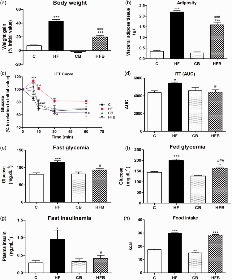
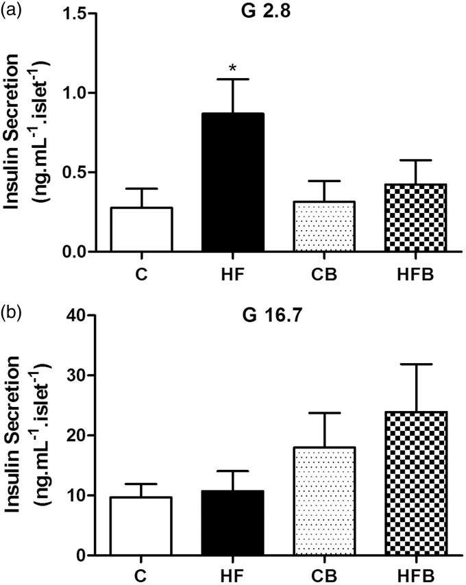
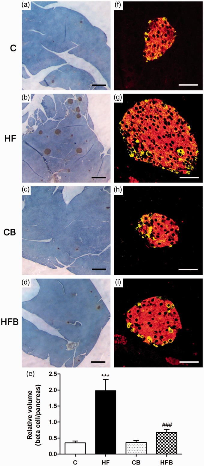
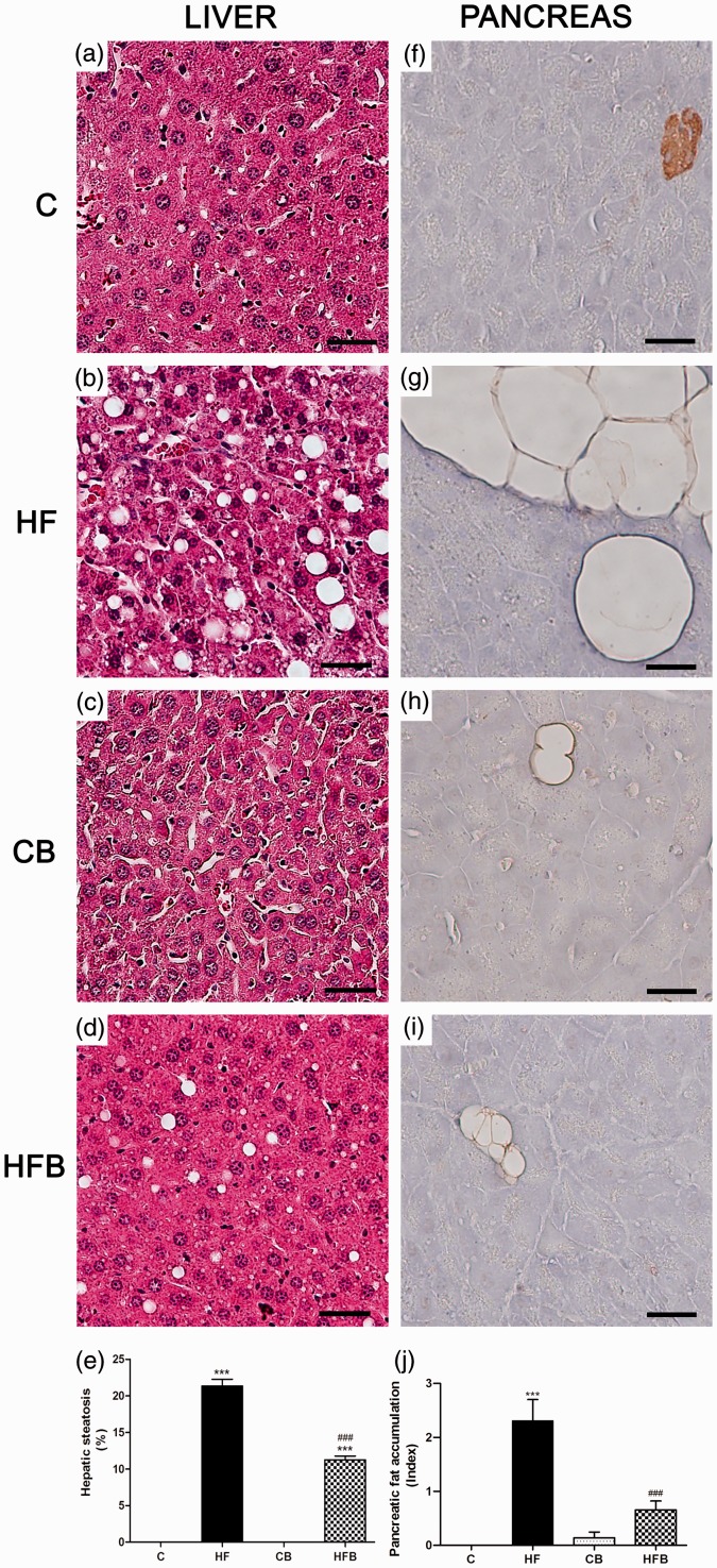
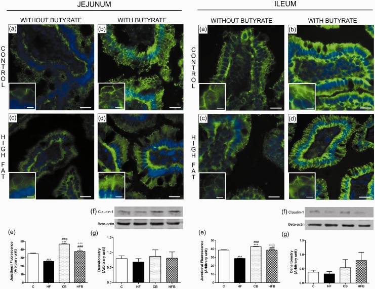
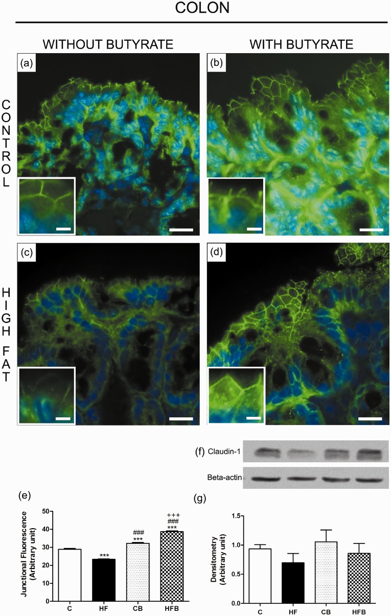
In this study, we investigated the effect of diet supplementation with sodium butyrate (5% w/w), a short-chain fatty acid produced by the intestinal microbiota, on metabolic parameters, body adiposity, hepatic and pancreatic lipid accumulation, beta cell function/mass as well as on the structure and function of the tight junction-mediated intestinal epithelial barrier in both normal and obese/prediabetic C57 mice fed a regular (control) or high-fat diet for 60 days, respectively. Butyrate treatment significantly inhibited all the high-fat-induced metabolic dysfunctions evaluated, i.e. significantly reduced the weight gain and body adiposity as well as the insulin resistant state, hyperglycemia and hyperinsulinemia, without changing food intake. In addition, high-fat-fed mice treated with this short-chain fatty acid displayed no compensatory hyperplasia of pancreatic beta cells nor marked hepatic steatosis as seen in prediabetic mice after high-fat diet only. Isolated pancreatic islets from high-fat-fed mice treated with butyrate showed improvement of the insulin secretion, which was associated with a significant decrease in lipid accumulation within the pancreas. Butyrate enhanced the intestinal epithelial barrier, as revealed by the FITC-Dextran permeability assay, which was accompanied by a significant increase in the junctional content of the tight junction-associated claudin-1 in intestinal epithelia of jejunum, ileum, and colon of both control and high-fat mice. In conclusion, our results showed that diet supplementation with butyrate inhibits the deleterious effects of high-fat diet intake on metabolic parameters and structure/function of several tissues/organs associated with type 2 diabetes mellitus in a mouse model, suggesting a potential use of this short-chain fatty acid in the treatment of this endocrine-metabolic disorder. Impact statement Butyrate is a short-chain fatty acid produced by the intestinal microbiota through the fermentation of non-absorbable carbohydrates and proteins (e.g. fibers). Sodium butyrate incorporated into the diet displayed a protective action on metabolic, hepatic, pancreatic and intestinal alterations induced by high-fat diet in mice, resulting in significant inhibition of the development of a prediabetic state. Thus, our data suggest that butyrate may have a potential therapeutic use in the treatment of type 2 diabetes and related disorders.

Keywords: Butyrate; high-fat diet; intestinal paracellular barrier; obesity; pancreatic beta cell; type 2 diabetes mellitus.


Figures

Figure 1
Figure 1
Butyrate treatment significantly reduced the high-fat diet-induced obesity and metabolic dysfunctions in mice without changing food intake. As compared to control group, high-fat (HF) diet for 60 days induced a significant increase in weight gain (a), adiposity (b), in peripheral insulin resistance (c), as assessed by the insulin tolerance test (ITT) and expressed as area values under the curve (AUC) (d), in fast (e) and fed (f) glycemia and in fast insulinemia (g), suggesting the development of prediabetes in these mice. However, dietary supplementation with 5% sodium butyrate (HFB group) showed a protective effect reducing significantly all these metabolic alterations induced by HF diet, without changing the food intake expressed as calories (h). Administration of butyrate per se (CB) did not significantly affect the parameters evaluated as compared to control group (C). Data are expressed as means + SE of at least three independent experiments (n = 14–18 mice per group). Groups: C = control (fed a regular diet alone); HF: high-fat diet alone; CB: Control + butyrate; HFB: high-fat diet + butyrate.*P < 0.05, **P < 0.001, ***P < 0.0001 compared to C group. #P < 0.05, ###P < 0.0001 compared to HF group. (A color version of this figure is available in the online journal.)
Figure 2
Figure 2
Pancreatic islets from high fat-fed mice show altered insulin secretion that was not observed after treatment with butyrate. Batches of isolated islets from mice of all experimental groups were exposed to 2.8 (a) or 16.7 mM glucose (b) for 60 min. Compared to controls (C), islets from HF-fed mice (HF) showed increased basal insulin secretion (expressed as ng.mL−1.islet−1) in the presence of 2.8 mM glucose (G 2.8) but a comparable insulin release when stimulated by 16.7 mM glucose (G 16.7) (b). This increased basal secretion was not observed in HF-fed mice receiving diet supplementation with butyrate (HFB) as compared to its control (CB)(a); but a tendency of an increase in stimulated insulin release (b) was verified in mice with butyrate diet supplementation (CB and HFD groups) as compared to those not receiving this SCFA (C and HF groups). Bars represent means + SE of four independent experiments (10–14 batches of five islets isolated from four mice per group). *P < 0.05 compared to C group
Figure 3
Figure 3
The compensatory beta cell mass expansion induced by high-fat diet in mice was not observed after butyrate administration. Photomicrographs of pancreatic islets processed for insulin immunoperoxidase (a–d) (brown) or for double immunofluorescence to detect insulin (red) and glucagon (yellow)(f–i). Note the increase in number and size of insulin-labelled islets (b,g), with no change in the islet cytoarchitecture (g), in high-fat diet-fed mice (HF) in comparison with control group (C) (a,f). This increase in the relative volume of insulin-producing beta cells induced by HF diet was not observed after butyrate treatment (HFB) (d,i) as compared to its control (CB) (c,h); this result was confirmed quantitatively as shown in (e). Bars in graph (e) represent means + SE of six mice per group. Images were obtained by light (a–d) or confocal laser microscopy (f–i). Scale bars, 500 µm in (a–d); 50 µm in (f–i). ***P < 0.0001 compared to C group; ###P < 0.0001 compared to HF group. (A color version of this figure is available in the online journal.)
Figure 4
Figure 4
Diet supplementation with butyrate inhibited hepatic steatosis and pancreatic fat accumulation induced by high-fat diet in mice. Photomicrographs of the liver (a–d) routinely processed for HE staining and pancreas processed for insulin immunoperoxidase (brown) (f–i). Exposure to high-fat (HF) diet-induced liver steatosis (b,e) and pancreatic fat accumulation (g,j) in HF mice as compared to controls (C) (a,f,e,j) that were significantly reduced by administration of butyrate (HFB) (d,i,e,j). No significant changes were observed with butyrate per se (CB group) (c,h) in comparison with C group (a,f). Scale bars, 50 µm. ***P < 0.0001 compared to C group ; ###P < 0.0001 compared to HF group. (A color version of this figure is available in the online journal.)
Figure 5
Figure 5
Butyrate, given as dietary supplementation, increased the junctional content of claudin-1, a tight junction-associated protein, in intestinal epithelia of the small intestine (jejunum and ileum). The junctional and total cell contents of claudin-1 were evaluated by immunofluorescence in intestinal cryosections (claudin-1 in green; DAPI/nuclei in blue) and by Western Blot in intestine homogenates, respectively. The analysis of the degree of fluorescence at the cell-cell contact showed a significant decrease in the intercellular content of claudin-1 in enterocytes of small intestine from HF diet-fed mice (HF group)(c,e), which was inhibited by the administration of butyrate to treated animals (HFB group) (d,e). The total cell content of this junctional protein displayed no significant changes after HF diet and/or butyrate treatment as revealed by immunoblotting (f,g). Groups: C: control (fed a regular diet alone); HF: high-fat diet alone; CB: control + butyrate; HFB: high-fat diet + butyrate. Scale bars, 15 µm (insets); 50 µm (images a–d).*P < 0.05, ***P < 0.0001 compared to C group, ###P < 0.0001 compared to HF group,+++P < 0.0001 compared to CB group. (A color version of this figure is available in the online journal.)
Figure 6
Figure 6
Butyrate inhibited the reduction in the junctional content of claudin-1 in enterocytes of the large intestine (colon) induced by exposure to the high-fat diet in mice. The junctional and total cell contents of claudin-1 were evaluated by immunofluorescence in colon cryosections (claudin-1 in green; DAPI/nuclei in blue) and by Western Blot in colon homogenates, respectively. The analysis of the degree of fluorescence at the cell–cell contact showed a significant decrease in the intercellular content of claudin-1 in enterocytes of colon from HF diet-fed mice (HF group)(c,e) as compared to controls (C group) (a,e); this was inhibited by the administration of butyrate to treated animals (HFB group) (d,e). The total cell content of this junctional protein displayed no significant changes after HF diet and/or butyrate treatment as revealed by immunoblotting (f,g). Groups: C: control (fed a regular diet alone); HF: high-fat diet alone; CB: control + butyrate; HFB: high-fat diet + butyrate. Scale bars, 15 µm (insets); 50 µm (images a–d). ***P < 0.0001 compared to the C group, ###P < 0.0001 compared to HF group, +++P < 0.0001 compared to CB group. (A color version of this figure is available in the online journal.)

References

    1. American Diabetes Association. Diagnosis and classification of diabetes mellitus. Diab Care 2014; 37: 81–90.
    1. Thomas CC, Philipson LH. Update on diabetes classification. Med Clin North Am 2015; 99: 1–16. - PubMed
    1. Canivell S, Gomis R. Diagnosis and classification of autoimmune diabetes mellitus. Autoimmun Rev 2014; 13: 403–7. - PubMed
    1. Olokoba AB, Obateru OA, Olokoba LB. Type 2 diabetes mellitus: a review of current trends. Oman Med J 2012; 27: 269–73. - PMC - PubMed
    1. Tripathy D, Chavez AO. Defects in insulin secretion and action in the pathogenesis of type 2 diabetes mellitus. Curr Diab Rep 2010; 10: 184–91. - PubMed

Publication types