. 2017 May 15;6(5):e337.
doi: 10.1038/oncsis.2017.36.

Molecular signaling in multiple myeloma: association of RAS/RAF mutations and MEK/ERK pathway activation

Affiliations

Molecular signaling in multiple myeloma: association of RAS/RAF mutations and MEK/ERK pathway activation

J Xu et al. Oncogenesis. .

Abstract

Multiple myeloma (MM) is a plasma cell malignancy that is still considered to be incurable in most cases. A dominant mutation cluster has been identified in RAS/RAF genes, emphasizing the potential significance of RAS/RAF/MEK/ERK signaling as a therapeutic target. As yet, however, the clinical relevance of this finding is unclear as clinical responses to MEK inhibition in RAS-mutant MM have been mixed. We therefore assessed RAS/RAF mutation status and MEK/ERK pathway activation by both targeted sequencing and phospho-ERK immunohistochemistry in 180 tissue biopsies from 103 patients with newly diagnosed MM (NDMM) and 77 patients with relapsed/refractory MM (rrMM). We found a significant enrichment of RAS/BRAF mutations in rrMM compared to NDMM (P=0.011), which was mainly due to an increase of NRAS mutations (P=0.010). As expected, BRAF mutations were significantly associated with activated downstream signaling. However, only KRAS and not NRAS mutations were associated with pathway activation compared to RAS/BRAFwt (P=0.030). More specifically, only KRASG12D and BRAFV600E were consistently associated with ERK activation (P<0.001 and P=0.006, respectively). Taken together, these results suggest the need for a more specific stratification strategy consisting of both confirmation of protein-level pathway activation as well as detailed RAS/RAF mutation status to allow for a more precise and more effective application of targeted therapies, for example, with BRAF/MEK inhibitors in MM.


Conflict of interest statement

The authors declare no conflict of interest.

Figures
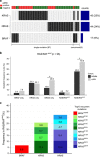
Figure 1
Figure 1
RAS/RAF mutation status and mutation frequency in MM. (a) A total of 96 out of 180 MM patients were identified with KRAS (45 cases), NRAS (44 cases) and BRAF mutations (17 cases) using a targeted panel (Ion AmpliSeq Cancer Hotspot panel v2, Ion Torrent/Thermo Fisher Scientific, Guilford, CT, USA), which covers KRAS/NRAS (exons 2, 3, 4), HRAS (exons 2, 3) and BRAF (exons 11,15). Targeted re-sequencing optimized for FFPE samples was performed as previously described., In brief, data were analyzed with the Ion Torrent Suite Software (version 3.6, Ion Torrent/Thermo Fisher Scientific) against reference human genome hg19 and annotated using the CLC Genomics Workbench (CLC Bio/Qiagen, Aarhus, Denmark, version 6.5) with integrated information about nucleotide and amino-acid changes from RefSeq annotated genes, COSMIC (version 69, COSMIC database, Wellcome Trust Sanger Institute, Cambridge, UK) and dbSNP databases. Only variants with a minimum coverage >200 reads were considered. About 1600 × mean coverage for each amplicon was achieved. Overall, RAS/RAF mutations exhibited a mutually exclusive pattern, with 90.6% of the RAS/RAFmut patients having single KRAS, NRAS or BRAF mutations, and only nine patients carried concurrent RAS/RAF mutations. The corresponding ERK activation status is shown on top, samples with moderate/strong immunohistochemical expression of pERK present in ⩾30% tumor cells were considered positive (red). (b) Comparison of RAS/RAF mutation frequencies in NDMM and rrMM cohorts. In general, RAS/RAF-mutant cases were significantly more frequent in rrMM compared to NDMM (*P<0.05). Notably, single NRAS mutations were significantly increased in rrMM, but not single BRAF or KRAS mutations. (c) Mutation frequencies in RAS/RAFmut samples of the 10 most recurrent non-synonymous mutations are shown as stacked bar plots grouped by genes. FFPE, formalin-fixed, paraffin-embedded; NDMM, newly diagnosed MM; pERK, phosphorylated ERK1/2; rrMM, relapsed/refractory MM; SNP, single-nucleotide polymorphism.
Figure 2
Figure 2
Evaluation of pERK expression in myeloma cells in bone marrow biopsies by IHC. The FFPE blocks were cut into 4–5 μm paraffin slides and stained with pERK antibody (#9101, Cell signaling, Danvers, MA, USA, 1:25, 24 min, 36 °C) using Ventana BenchMark Ultra Autostainer (Ventana Medical Systems/Roche, Tucson, AZ, USA) and OptiView DAB IHC detection Kit (Ventana Medical Systems/Roche). The staining protocol was optimized for pERK with CC1 (pH8.4) pretreatment for 64 min. For further information on effects of preservation protocols and controls, please refer to Supplementary Figure S2. The intensity of cytoplasmic staining of pERK in myeloma cells (i) was assessed in relation to endothelial cells (internal positive control cells for pERK, depicted by red arrow heads) and categorized into negative, low, moderate and strong (score 0–3), the percentage of positive tumor cells (N) was scored from 0 to 10, representing 0–100% of total assessable tumor cells. The cases with moderate/strong immunohistochemical expression of pERK present in ⩾30% tumor cells were classified as positive. FFPE, formalin-fixed, paraffin-embedded; IHC, immunohistochemistry; pERK, phosphorylated ERK1/2.
Figure 3
Figure 3
Correlation of RAS/RAF mutations with pERK expression. (a) BRAFmut, KRASmut and NRASmut cases were compared to RAS/BRAFwt cases in relation to ERK activation. (b) Cases harboring one of the 10 most recurrent RAS/RAF mutations were tested against all RAS/RAFwt cases for ERK activation status. A consistent association of ERK activation was only observed in cases with KRASG12D (P<0.001) and BRAFV600E (P=0.006). AllP-values were calculated using Fisher’s exact test. pERK, phosphorylated ERK1/2.

References

    1. Bolli N, Avet-Loiseau H, Wedge DC, Van Loo P, Alexandrov LB, Martincorena I et al. Heterogeneity of genomic evolution and mutational profiles in multiple myeloma. Nat Commun 2014; 5: 2997. - PMC - PubMed
    1. Lohr JG, Stojanov P, Carter SL, Cruz-Gordillo P, Lawrence MS, Auclair D et al. Widespread genetic heterogeneity in multiple myeloma: implications for targeted therapy. Cancer Cell 2014; 25: 91–101. - PMC - PubMed
    1. Walker BA, Boyle EM, Wardell CP, Murison A, Begum DB, Dahir NM et al. Mutational spectrum, copy number changes, and outcome: results of a sequencing study of patients with newly diagnosed myeloma. J Clin Oncol 2015; 33: 3911–3920. - PMC - PubMed
    1. Heuck CJ, Jethava Y, Khan R, van Rhee F, Zangari M, Chavan S et al. Inhibiting MEK in MAPK pathway-activated myeloma. Leukemia 2015; 30: 976–980. - PMC - PubMed
    1. Mandal R, Becker S, Strebhardt K. Stamping out RAF and MEK1/2 to inhibit the ERK1/2 pathway: an emerging threat to anticancer therapy. Oncogene 2016; 35: 2547–2561. - PubMed