

H3F3A (Histone 3.3) G34W Immunohistochemistry: A Reliable Marker Defining Benign and Malignant Giant Cell Tumor of Bone
- PMID: 28505000
- PMCID: PMC5510691
- DOI: 10.1097/PAS.0000000000000859
H3F3A (Histone 3.3) G34W Immunohistochemistry: A Reliable Marker Defining Benign and Malignant Giant Cell Tumor of Bone
Abstract
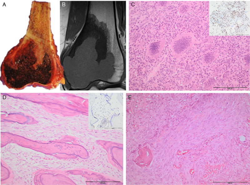
Giant cell tumor of bone (GCTB) is a locally aggressive subarticular tumor. Having recently reported that H3.3 G34W mutations are characteristic of this tumor type, we have now investigated the sensitivity and specificity of the anti-histone H3.3 G34W rabbit monoclonal antibody in a wide variety of tumors including histologic mimics of GCTB to assess its value as a diagnostic marker. We also determined the incidence of H3.3 G34 mutations in primary malignant bone tumors as assessed by genotype and H3.3 G34W immunostaining. A total of 3163 tumors were tested. Totally, 213/235 GCTB (90.6%) showed nuclear H3.3 p.G34W immunoreactivity. This was not the case for the rare variants, p.G34L, M, and V, which occurred most commonly in the small bones of the hands, patella, and the axial skeleton. If these sites were excluded from the analysis, H3.3 G34W expression was found in 97.8% of GCTB. Malignant bone tumors initially classified as osteosarcomas were the only other lesions (n=11) that showed G34W expression. Notably an additional 2 previously reported osteosarcomas with a p.G34R mutation were not immunoreactive for the antibody. A total of 11/13 of these malignant H3.3-mutant tumors exhibited an osteoclast-rich component: when imaging was available all but one presented at a subarticular site. We propose that subarticular primary malignant bone sarcoma with H3.3 mutations represent true malignant GCTB, even in the absence of a benign GCTB component.
Conflict of interest statement
Conflicts of Interest and Source of Funding: Supported by the Medical Research Council grant (MR/M00094X/1). A grant awarded to A.M.F. by the Royal National Orthopaedic Hospital NHS Trust NHS Trust, and the Skeletal Cancer Action Trust (Scat), UK. A.M.F. was supported by the National Institute for Health Research, UCLH Biomedical Research Centre, and the UCL Experimental Cancer Centre. D.B. was supported by the Gertrude von Meissner-Stiftung and the Basel Bone Tumour Reference Centre Foundation. The authors have disclosed that they have no significant relationships with, or financial interest in, any commercial companies pertaining to this article.
Figures


References
-
- Athanasou NA, Bansal M, Forsyth R, et al. Fletcher C, Bridge J, Hogendoorn P, et al. Giant cell tumour of bone. WHO Classification of Tumours of Soft Tissue and Bone. Lyon, France: IARC Press; 2013:321–324.
-
- Errani C, Ruggieri P, Asenzio MA, et al. Giant cell tumor of the extremity: a review of 349 cases from a single institution. Cancer Treat Rev. 2010;36:1–7. - PubMed
-
- Forsyth R, Jundt G.Fletcher CDM, Bridge JA, Hogendoorn PCW, et al. Giant cell lesion of the small bones. WHO Classification of Tumours of Soft Tissue and Bone. Lyon, France: IARC Press; 2013:320.
-
- Al-Ibraheemi A, Inwards CY, Zreik RT, et al. Histologic spectrum of giant cell tumor (GCT) of bone in patients 18 years of age and below: a study of 63 patients. Am J Surg Pathol. 2016;40:1702–1712. - PubMed
MeSH terms
Substances
Grants and funding
LinkOut - more resources
Full Text Sources
Other Literature Sources
Medical
