. 2017 May 15;18(5):1061.
doi: 10.3390/ijms18051061.

Upregulated Autophagy in Sertoli Cells of Ethanol-Treated Rats Is Associated with Induction of Inducible Nitric Oxide Synthase (iNOS), Androgen Receptor Suppression and Germ Cell Apoptosis

Affiliations

Upregulated Autophagy in Sertoli Cells of Ethanol-Treated Rats Is Associated with Induction of Inducible Nitric Oxide Synthase (iNOS), Androgen Receptor Suppression and Germ Cell Apoptosis

Akio Horibe et al. Int J Mol Sci. .

Abstract

This study was conducted to investigate the autophagic response of Sertoli cells (SCs) to acute ethanol toxicity using in vivo and in vitro models. Adult Wistar rats were intraperitoneally injected with either 5 g/kg ethanol or phosphate-buffered saline (for the control group) and sacrificed 0, 3, 6 and 24 h after injection. Compared to the control group, enhanced germ cell apoptosis was observed in the ethanol-treated rats (ETRs) in association with upregulation of iNOS and reduced expression of androgen receptor protein levels in SCs, which were resistant to apoptosis. Meanwhile, autophagy was upregulated in ETR SCs (peaking at 24 h) compared to the control group, as evidenced by transcription factor EB (TFEB) nuclear translocation, enhanced expression of microtubule-associated protein 1 light chain3-II (LC3-II), lysosome-associated membrane protein-2 (LAMP-2), pan cathepsin protein levels and reduced expression of p62. This upregulation of SC autophagy was confirmed ultrastructurally by enhanced formation of autophagic vacuoles and by immunofluorescent double labelling of autophagosomal and lysosomal markers. Study of cultured SCs confirmed enhanced autophagic response to ethanol toxicity, which was cytoprotective based on decreased viability of SCs upon blocking autophagy with 3-methyladenine (3-MA). The results highlighted the molecular mechanisms of prosurvival autophagy in ETR SCs for the first time, and may have significant implications for male fertility.

Keywords: Sertoli; androgen receptor (AR); apoptosis; autophagy; ethanol; inducible nitric oxide synthase (iNOS); transcription factor EB (TFEB).


Conflict of interest statement

The authors declare no conflict of interest.

Figures
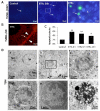
Figure 1
Figure 1
Ethanol-enhanced germ cell apoptosis. (A) Terminal deoxynucleotidyl transferase dUTP-mediated nick-end labelling (TUNEL) labelling of apoptotic germ cells (green reaction, white arrows). 4′,6-diamidino-2-phenylindole (DAPI) (blue staining) is used for nuclear counterstaining; (B) TUNEL/AR (SC marker) double labelling. The arrow heads mark nuclei of SCs expressing AR (red labelling); (C) Histogram showing significant increase of the percentage of apoptotic STs in ETRs. * p < 0.05; ** p < 0.01; (D) TEM demonstrating normal germ cells in control testis (a) and apoptotic germ cells in ETRs (bf). The framed area in b is magnified in c. S: SC nucleus; Spg: spermatogonia; Sp: spermatid; Spr: spermatocyte; AR: androgen receptor; SCs: Sertoli cells; STs: seminiferous tubules; ETRs: ethanol-treated rats; TEM: transmission electron microscopy. Scale bars in A, B: 50 μm for first two panels; 20 μm for next two panels.
Figure 2
Figure 2
Upregulation of inducible nitric oxide synthase (iNOS) and suppression of ARs in SCs and interstitial cells of ETRs. (A,B) show the immunohistochemistry (IHC) of iNOS, while (C,D) demonstrate the IHC of AR. The framed areas in (A,C) are magnified in (B,D). The black arrows in (A,B) mark iNOS expression in SCs, and the red arrow shows its expression in an interstitial cell. Black, red and green arrow heads in (C,D) indicate nuclear expression of AR in SCs, Leydig and myoid cells, respectively. Scale bars in A, C: 20 μm.
Figure 3
Figure 3
Induction and upregulation of the autophagy marker microtubule-associated protein 1 light chain 3 (LC3) in ETR SCs. (A) toluidine blue staining of semi-thin sections demonstrating perinuclear vacuole formation (black arrows) and lipid droplet (yellow arrows) accumulation in ETR SCs. The framed areas are magnified in the insets; (B,C) show immunofluorescence (IF) and IHC of LC3 expression, respectively. S: SC nucleus; (D) shows quantification of LC3 puncta. The boxed areas in B are magnified on the right. The red arrows mark LC3 puncta in SCs. * p < 0.05. The scale bar for left panels in B is 20 μm.
Figure 4
Figure 4
Elevated colocalization of LC3 with lysosomal cathepsins in ETR SCs, indicating enhanced autophagic activity. IF double labelling of LC3 (red) and pan cathepsin (pan c) (green) in control and ETR testes (a). The colocalization signals observed upon merging (yellow-orange) (b) are marked by yellow arrows (c) and shown as white spots (d) using Image J. S: SC nucleus. The histograms (e) are plot profiles indicating the overexpression and colocalization of LC3 (red) and pan cathepsin (green) in SCs, and correlate to the white lines shown in the images. The scale bar in b is 20 μm. The panels in a and ce are lower and higher magnifications of a, respectively using image J.
Figure 5
Figure 5
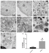
Ultrastructural features of upregulated autophagy in ETR SCs. TEM of control (A) and ETRs (BG). The histogram (H) shows a significant increase in the number of autophagic vacuoles (AVs) in ETR SCs. The long black arrows mark autophagosomes with a double limiting membrane (arrow heads) (magnified in the inset in (C). The short arrows indicate autolysosomes. The broken arrows in (E) show multilamellar bodies, while the white arrows in (G) show autophagosomes containing fragmented mitochondria. S: SC nucleus; L: lysosome; M: mitochondria; LD: lipid droplet. * p < 0.05.
Figure 6
Figure 6
Upregulation and transcription factor EB (TFEB) nuclear translocation in ETR SCs are associated with increased expression of lysosomal proteins. (A,B) show IHC of TFEB and pan cathepsin, respectively. The framed SC nuclei in (A) are magnified in the insets, and arrow heads show nuclear expression of TFEB. The framed areas of SCs in (B) are magnified in the insets. Black arrows show pan cathepsin expression in SCs.
Figure 7
Figure 7
Ethanol (EtOH) enhanced autophagy in cultured SCs as an apparent prosurvival mechanism. (A,B) show LC3 staining and quantification, and (C) shows western blot of LC3-II. The framed areas in A are magnified in the insets. The white arrows indicate LC3 puncta; (D) shows a histogram indicating reduced viability of SCs on autophagy blocking with 3-methyladenine (3-MA). * p < 0.05; ** & ## p < 0.01 significantly different from the corresponding group. The statistical analysis of western blot is based on five independent experiments.
Figure 8
Figure 8
A schema showing the molecular mechanisms, evidences and consequences of ethanol-induced upregulation of autophagy in SCs.

References

    1. França L.R., Hess R.A., Dufour J.M., Hofmann M.C., Griswold M.D. The Sertoli cell: One hundred fifty years of beauty and plasticity. Andrology. 2016;4:189–212. doi: 10.1111/andr.12165. - DOI - PMC - PubMed
    1. Rato L., Meneses M.J, Silva B.M., Sousa M., Alves M.G., Oliveira P.F. New insights on hormones and factors that modulate Sertoli cell metabolism. Histol. Histopathol. 2016;31:499–513. - PubMed
    1. Oliveira P.F., Martins A.D., Moreira A.C., Cheng C.Y., Alves M.G. The Warburg effect revisited-lesson from the Sertoli cell. Med. Res. Rev. 2015;35:126–151. doi: 10.1002/med.21325. - DOI - PMC - PubMed
    1. Lejeune H., Skalli M., Chatelain P.G., Avallet O., Saez J.M. The paracrine role of Sertoli cells on Leydig cell function. Cell Biol. Toxicol. 1992;8:73–83. doi: 10.1007/BF00130513. - DOI - PubMed
    1. Sanberg P.R., Borlongan C.V., Othberg A., Saporta S., Freeman T.B., Cameron D.F. Testis-derived Sertoli cells have a trophic effect on dopamine neurons and alleviate hemiparkinsonism in rats. Nat. Med. 1997;3:1129–1132. doi: 10.1038/nm1097-1129. - DOI - PubMed

MeSH terms

LinkOut - more resources