. 2017 Aug 15;26(16):3081-3093.
doi: 10.1093/hmg/ddx192.

Sialic acid deficiency is associated with oxidative stress leading to muscle atrophy and weakness in GNE myopathy

Affiliations

Sialic acid deficiency is associated with oxidative stress leading to muscle atrophy and weakness in GNE myopathy

Anna Cho et al. Hum Mol Genet. .

Abstract

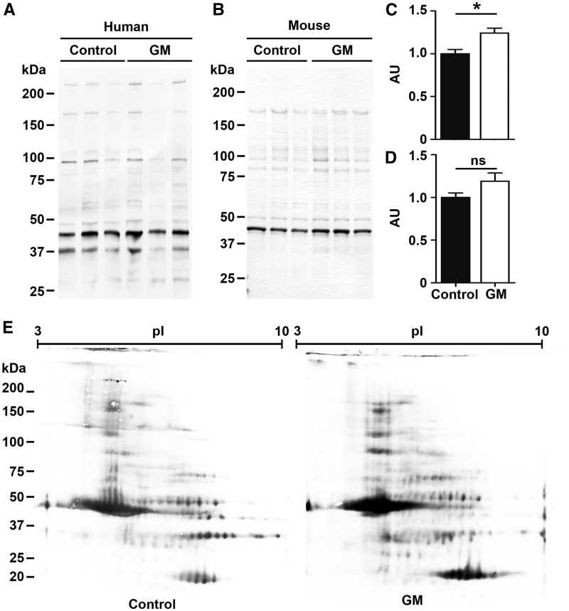
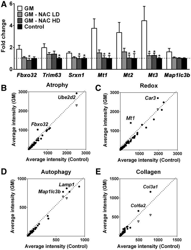
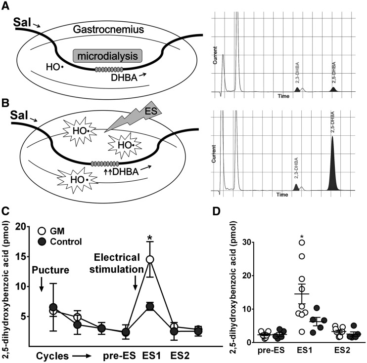
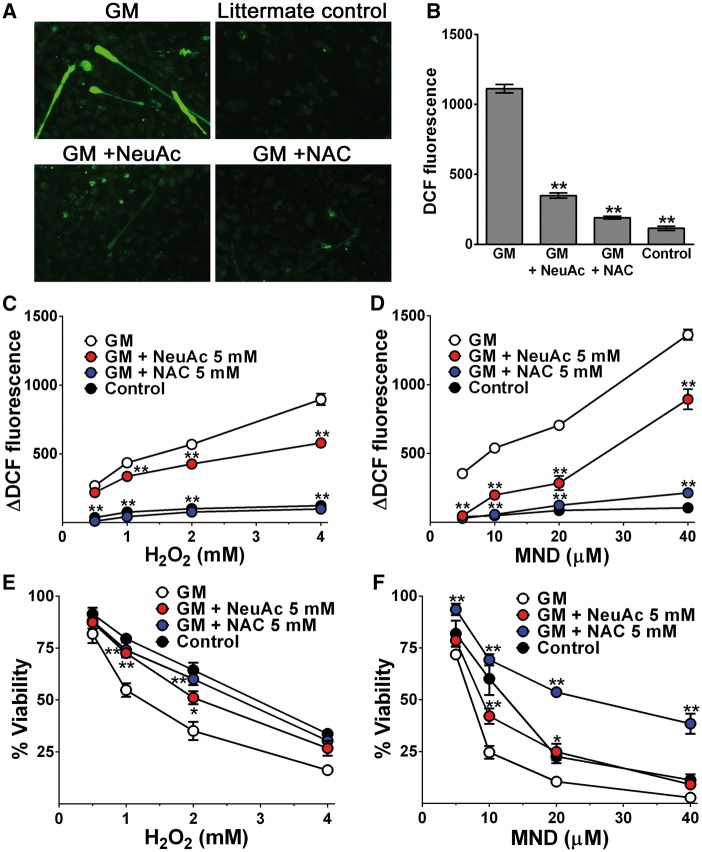
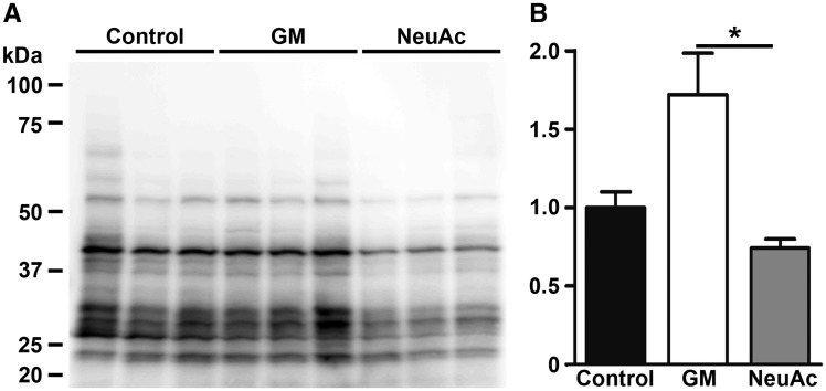
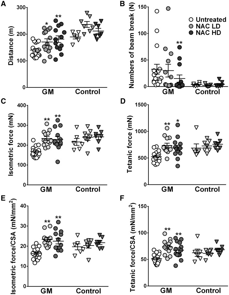
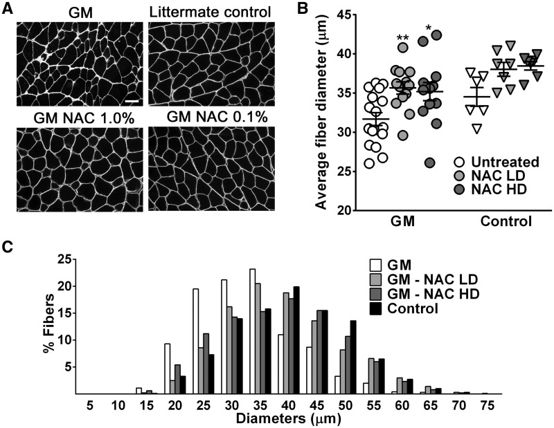
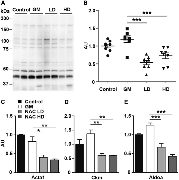
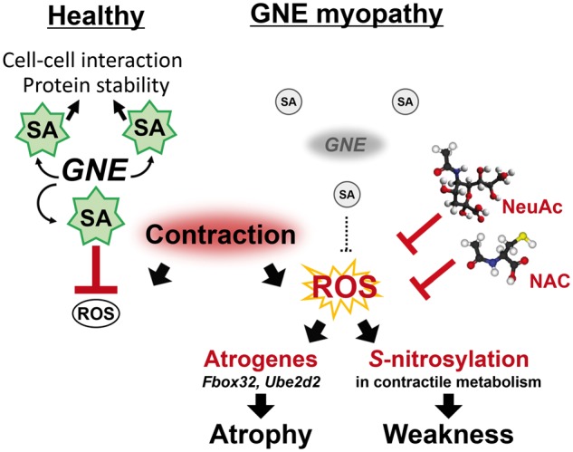
Sialic acids are monosaccharides found in terminal sugar chains of cell surfaces and proteins; they have various biological functions and have been implicated in health and disease. Genetic defects of the GNE gene which encodes a critical bifunctional enzyme for sialic acid biosynthesis, lead to GNE myopathy, a disease manifesting with progressive muscle atrophy and weakness. The likely mechanism of disease is a lack of sialic acids. There remains, however, an unexplained link between hyposialylation and the muscle atrophy and weakness. In this study, we found that muscle proteins were highly modified by S-nitrosylation, and that oxidative stress-responsive genes were significantly upregulated, in hyposialylated muscles from human GNE myopathy patients and model mice. In both in vitro and in vivo models, the production of reactive oxygen species (ROS) was elevated with cellular hyposialylation, and increasing overall sialylation by extrinsic sialic acid intake reduced ROS and protein S-nitrosylation. More importantly, the antioxidant, oral N-acetylcysteine led to amelioration of the muscle atrophy and weakness in Gne mutant mice. Our data provide evidence of additional important function of sialic acids as a ROS scavenger in skeletal muscles, expanding our understanding on how sialic acid deficiency contributes to disease pathology, and identify oxidative stress as a therapeutic target in GNE myopathy.


Figures

Figure 1
Figure 1
Elevated S-nitrosylation of skeletal muscle proteins in GNE myopathy muscles. (A) Detection of protein S-nitrosylation in human skeletal muscles. Control, diseased control; GM, GNE myopathy patients (left, Patient 1; center, Patient 2; right, Patient 3). (B) Detection of S-nitrosylated proteins from mouse skeletal muscles. Control, littermates; GM, GNE myopathy mice. (C) Total protein S-nitrosylation rates in human GNE myopathy muscles (n = 4). (D) Total S-nitrosylation rates in mouse muscle proteins (n = 7). Total intensities were normalized by total protein amounts. Data represent mean ± SEM of each group. *P < 0.05. (E)S-nitrosylated protein profiles on 2-Dimensional PAGE. GM, GNE myopathy patient 5.
Figure 2
Figure 2
Upregulation of atrogenes and oxidative stress-responsive genes in GNE myopathy muscles. (A) Expression of Fbxo32, Trim63, Srxn1, Mts, and Map1lc3b in untreated (white bars, n = 17), high dose (HD, 1.0%) or low dose (LD, 0.1%) N-acetylcysteine (NAC) treated (gray bars, n = 6 per group), and littermate controls (black bars, n = 6) as fold changes of littermate controls. Data represent mean ± SEM of each group. *P < 0.05. (B-E) Microarray analysis followed by gene ontology profiling: (B) muscle atrophy-related genes, (C) oxidative stress and redox homeostasis-related genes, (D) autophagy-related genes, (E) collagen-related genes. Each dot represents average expression values for the same gene from GNE myopathy (vertical axis, n = 9) and littermates (horizontal axis, n = 3) muscles. Inverted triangles show recovered expression values with N-acetylcysteine treatment (vertical axis, n = 6) for significantly upregulated genes.
Figure 3
Figure 3
Measurement of ROS production in the GNE myopathy muscle in vivo using microdialysis. (A-B) Experimental set up and representative HPLC chromatograms. Perfusion medium containing 5 mM salicylate (Sal) was pumped through a microdialysis probe and DHBA in dialysate was detected by HPLC–electrochemical detection system: (A) baseline, (B) post-stimulus. (C) Electrical stimulus (ES) consisted of 50V, 40 Hz, 3 ms pulses for 300 trains. HPLC profils of DHBA during the experiments. (D) DHBA increments after contraction in GNE myopathy mice (n = 9) and littermates (n = 5). Pre-ES, before ES. ES1, 1st 20 min after ES. ES2, 2nd 20 min after ES. Data represent mean ± SEM. Each circle represents a DHBA level from an individual mouse. *P < 0.05.
Figure 4
Figure 4
Impaired antioxidant capacity in hyposialylated GNE myopathy myotubes. (A) Intracellular ROS generation with green DCF staining. (B) Measurement of DCF fluorescence by a plate reader. Data represent mean ± SEM of each group (n = 14). (C-D) ROS levels and cells viability were analysed with the addition of increasing concentration of H2O2 (0.5, 1.0, 2.0, and 4.0 mM) or menadione (5, 10, 20, and 40 μM) to culture media. Control myotubes of each group were cultured in the same condition without adding H2O2, menadione: (C) increased ROS levels with the addition of increasing concentration of H2O2, (D) menadione. (E-F) Relative viability of cells: (E) with the addition of increasing concentration of H2O2, (F) menadione. N-acetylcysteine (NAC). Each point is mean ± SEM of four determinations. *P < 0.05; **P < 0.01.
Figure 5
Figure 5
Suppression of protein S-nitrosylation in GNE myopathy mice muscles by sialic acid administration. (A,B) Protein S-nitrosyl modification in gastrocnemius muscles from the GNE myopathy mice with/without oral administration of NeuAc at 20 mg/kg body wt/d for 10 mo. Control, untreated controls (n = 3); GM, untreated GNE myopathy mice (n = 3); NeuAc NeuAc-treated GNE myopathy mice (n = 3). Data presented with mean ± SEM. *P < 0.05.
Figure 6
Figure 6
Oral N-acetylcysteine administration improved muscle force generation and motor performance in GNE myopathy mice. GNE myopathy mice were treated with low dose (LD, n = 13) or high dose (HD, n = 13) N-acetylcysteine (NAC) and compared to untreated group (n = 17). Littermate controls were treated in the same conditions (LD, n = 7; HD, n = 7; untreated, n = 6). (A) Treadmil performance test evaluating the distance mice could run. (B) Treadmill endurace test evaluating the number of beam breaks during a constant loading. (C-F) Contractile forces of gastrocnemius muscles: (C) in peak isometric twitch force, (D) maximum tetanic force, (E) specific isometric force normalized by CSA, (F) specific tetanic force normalized by CSA. Values from each mouse are shown with mean ± SEM. *P < 0.05; **P < 0.01.
Figure 7
Figure 7
Skeletal muscle atrophy in GNE myopathy mice was ameliorated by N-acetylcysteine treatment. (A) Representative sarcolemmal staining images with caveolin-3 antibody from gastrocnemius muscles. NAC, N-acetylcysteine treated mice. Bar = 20 μm. (B) Muscle fiber diameters in low dose (LD, n = 13) or high dose (HD, n = 13) N-acetylcysteine treated GNE myopathy mice (NACLD or NACHD) were compared to those in untreated controls (n = 17). Data presented with mean ± SEM. *P < 0.05; **P < 0.01. (C) Fiber diameter histogram from a mouse in each group was compared.
Figure 8
Figure 8
Reduction of S-nitrosyl modification in skeletal muscle proteins by N-acetylcysteine treatment. (A) Profiles of S-nitrosylation of mouse skeletal muscle proteins. (B) Total S-nitrosylation rates nomalized by protein amounts. (C–E)S-nitrosylation rates of skeletal muscle: (C) actin (Acta1), (D) muscle creatine kinase (Ckm), (E) fructose-bisphosphate aldolase A (Aldoa). Control, untreated controls (n = 8); GM, untreated GNE myopathy mice (n = 7); LD/NACLD, GM with low dose of N-acetylcysteine (n = 8); HD/NACHD, GM with high dose of N-acetylcystine (n = 8). Data presented with mean ± SEM. *P < 0.05; **P < 0.01; ***P < 0.001.
Figure 9
Figure 9
Role of sialic acids on antioxidant defense system. Left, sialic acids (SA) have a role to scavenge ROS generated on muscle contraction in healthy skeletal muscles. Right, proposed pathomechanism in GNE myopathy. Muscle atrophy and weakness are promoted by enhanced oxidative stress in reduced SA in skeletal muscles due to GNE mutations. Extrinsic NeuAc or antioxidant (NAC, N-acetylcysteine) supplementation has an effect against myopathic phenotypes in GNE myopathy model mice.

References

    1. Pillai S., Netravali I.A., Cariappa A., Mattoo H. (2012) Siglecs and immune regulation. Annu. Rev. Immunol., 30, 357–392. - PMC - PubMed
    1. Jaeken J. (2013) Congenital disorders of glycosylation. Handb. Clin. Neurol., 113, 1737–1743. - PubMed
    1. Schnaar R.L., Gerardy-Schahn R., Hildebrandt H. (2014) Sialic acids in the brain: gangliosides and polysialic acid in nervous system development, stability, disease, and regeneration. Physiol. Rev., 94, 461–518. - PMC - PubMed
    1. Keppler O.T., Hinderlich S., Langner J., Schwartz-Albiez R., Reutter W., Pawlita M. (1999) UDP-GlcNAc 2-epimerase: a regulator of cell surface sialylation. Science, 284, 1372–1376. - PubMed
    1. Seppala R., Lehto V.P., Gahl W.A. (1999) Mutations in the human UDP-N-acetylglucosamine 2-epimerase gene define the disease sialuria and the allosteric site of the enzyme. Am. J. Hum. Genet., 64, 1563–1569. - PMC - PubMed

Publication types

MeSH terms

Substances