. 2017 Jul 27;45(13):8064-8078.
doi: 10.1093/nar/gkx419.

The herpes viral transcription factor ICP4 forms a novel DNA recognition complex

Affiliations

The herpes viral transcription factor ICP4 forms a novel DNA recognition complex

Richard B Tunnicliffe et al. Nucleic Acids Res. .

Abstract

The transcription factor ICP4 from herpes simplex virus has a central role in regulating the gene expression cascade which controls viral infection. Here we present the crystal structure of the functionally essential ICP4 DNA binding domain in complex with a segment from its own promoter, revealing a novel homo-dimeric fold. We also studied the complex in solution by small angle X-Ray scattering, nuclear magnetic resonance and surface-plasmon resonance which indicated that, in addition to the globular domain, a flanking intrinsically disordered region also recognizes DNA. Together the data provides a rationale for the bi-partite nature of the ICP4 DNA recognition consensus sequence as the globular and disordered regions bind synergistically to adjacent DNA motifs. Therefore in common with its eukaryotic host, the viral transcription factor ICP4 utilizes disordered regions to enhance the affinity and tune the specificity of DNA interactions in tandem with a globular domain.


Figures

Figure 1.
Figure 1.
Summary of ICP4 domains and sites of DNA interaction and homo-dimerization. (A) The domain composition of HSV1 ICP4 with predicted secondary structure (SSpred) and ordered regions (ORDERpred) from PSIPRED (37). Predicted α-helices and β-sheets are shown as red and yellow respectively, with ordered regions colored green. (B) Sequence alignment of ICP4 DBDs from HSV1, HSV2 and VZV (Uniprot codes: P08392, P90493, Q8AZM1 respectively) from Clustal omega (36). Secondary structure elements determined here are labeled on the HSV-1 sequence. Residues previously probed by mutagenesis are highlighted blue with black arrows pointing to those with lower affinity for DNA (13,14,16). Below the sequences, blocks colored gray or purple indicate residues which form homo-dimer or DNA contacts respectively as observed in the crystallography data. Purple circles indicate protein–DNA contacts derived from NMR chemical shift perturbations in intrinsically disordered regions (IDRs). (C) Cartoon representation of the crystal structure with helices, sheets and loops shown as cylinders, arrows and coil respectively: (i) ICP4N·IE3_19mer with DNA colored purple and pink for sense and anti-sense strands respectively, one protein chain is colored gray and other blue through red from N-to-C termini. (ii) As with panel i with view rotated 90°.
Figure 2.
Figure 2.
Structural details of and biophysical characterization of ICP4N dimerization. (A) Cartoon of protein chains C and D (colored blue and green respectively) from the ICP4N·IE3_19mer structure with α-helices 5, 6 and 7 highlighted which form the major hydrophobic homo-dimer interface. (B) Details of the residues within the major homo-dimer interface, hydrophobic contacts are indicated by orange lines and hydrogen bonds by pink dashes. (C) SEC-MALS profile of ICP4N with and without IE3_19mer DNA, shown as dashed or solid lines respectively and refractive index (black lines) and molecular mass (green lines), plotted against elution volume. (D) Velocity AUC analysis of ICP4N with and without IE3_19mer DNA, shown as dashed or solid lines respectively. For each sample a major peak was observed corresponding to a dimeric protein, free or in complex with DNA.
Figure 3.
Figure 3.
Details of the protein–DNA interface in the ICP4N·IE3_19mer structure. (A) Schematic of ICP4N-IE3 DNA interaction model. Protein–DNA hydrogen bonds identified from the crystal structure are indicated by dashed lines with locations of other contacts indicated with dark gray lines. Protein residues are colored blue or green when corresponding to chain C or D respectively. The vertical dashed arrow marks the DNA region that NMR, SAXS and SPR data suggested is bound by an IDR of the protein (residues 258–289). ICP4 consensus is sequence shown to the left side. (B) Overall ICP4N·IE3_19mer structure, with protein chains C and colored blue and green respectively and DNA space fill surface is shown colored dark and light orange for the sense (chain H) and antisense strands (chain G) respectively. Hydrogen bonds are indicated by dashes. Details of base-pair interactions, shown for (i) major groove bound by the ICP4N globular homo-dimer residues 416–419 and 456–457, (ii) DNA kink intercalated by F283 and (iii) minor groove bound by residues 416–419 and 456–457. (C) Surface of the ICP4N dimer colored by electrostatic potential (red through blue for acidic to basic charge) calculated by Adaptive Poisson-Boltzmann Solver module in Chimera (53). DNA is shown in cartoon form with the ICP4 consensus sequence labeled on sense strand. (D) Plot of DNA major- and minor-groove widths measured in both models in the ICP4N·IE3_19mer asymmetric unit (54).
Figure 4.
Figure 4.
SAXS and NMR analysis of the ICP4N·IE3_19mer complex. (A) Dimensionless Kratky plot of the ICP4N bound (red) and unbound (blue) to DNA. Cross-hairs denote the Guinier–Kratky point (1.732, 1.104), the peak position for an ideal, globular particle. As indicated by the upward-right shift of the peaks in the dimensionless Kratky plot, ICP4N is more globular in the presence of DNA. (B) The calculated solution-state SAXS profile for the crystal structures of ICP4N·IE3_19mer complex (black line) compared to the measured scatter data (red circles). (C) Multi-phase ab initio model generated from SAXS data using MONSA show the presence of DNA (orange) and protein (teal) and their arrangement. (D) The crystal structure of the complex docked into the ab initio model revealing unoccupied volume around the DNA as well as above and below the protein dimer, assigned to the N-terminal IDRs. (E) NMR characterization of IDRs of the ICP4N dimer upon addition of equimolar amount of IE3_19mer duplex. 1H-15N TROSY spectrum of ICP4N showing sharp peaks assigned to residues within the unstructured N- and C-terminal regions in free and IE3 DNA bound forms, colored blue and red respectively. Peaks are labeled with assignments; when an unambiguous assignment was not possible the peaks are labeled with their amino acid type.
Figure 5.
Figure 5.
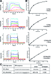
Binding of ICP4N and ICP4ΔIDR to biotinylated DNA duplexes measured by SPR. (A) Sensorgrams of different concentrations (nano-molar concentrations indicated on each plot) of ICP4 proteins binding to IE3 DNA duplexes. (B) Equilibrium analysis of SPR. (C) Mean dissociation constants (±SD) measured for each interaction, non-WT bases are underlined.
Figure 6.
Figure 6.
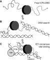
Model of action of the ICP4 DNA binding domain (DBD). The globular homo-dimer is represented by gray and black ovals and N-terminal IDRs by lines. (A) Free protein adopts an expanded conformation with the IDRs extended. (B) When not in contact with a DNA consensus site, ICP4-DBD and particularly the IDRs search DNA strands for sequence motifs. (C) Binding to the IE3 consensus site which overlaps with the transcription initiation site, ICP4 forms an asymmetric complex by the synergistic action of the globular region to the RTCGTC motif and an IDR with the downstream YnYSG motif. One IDR is not involved in specific DNA recognition and points upstream toward the TATA box, which is compatible with tripartite complex formation by the TATA binding protein, TFIIB and ICP4.

Similar articles

Cited by

References

    1. Itzhaki R.F. Herpes simplex virus type 1 and Alzheimer's disease: increasing evidence for a major role of the virus. Front. Aging Neurosci. 2014; 6:202. - PMC - PubMed
    1. Manservigi R., Argnani R., Marconi P.. HSV recombinant vectors for gene therapy. Open Virol. J. 2010; 4:123–156. - PMC - PubMed
    1. Russell S.J., Peng K.W., Bell J.C.. Oncolytic virotherapy. Nat. Biotechnol. 2012; 30:658–670. - PMC - PubMed
    1. Honess R.W., Roizman B.. Regulation of herpesvirus macromolecular synthesis: sequential transition of polypeptide synthesis requires functional viral polypeptides. Proc. Natl. Acad. Sci. U.S.A. 1975; 72:1276–1280. - PMC - PubMed
    1. Gruffat H., Marchione R., Manet E.. Herpesvirus late gene expression: a viral-specific pre-initiation complex is key. Front. Microbiol. 2016; 7:869. - PMC - PubMed

MeSH terms

Substances