. 2017 Sep 1;12(9):1402-1413.
doi: 10.1093/scan/nsx067.

Potential reward reduces the adverse impact of negative distractor stimuli

Affiliations

Potential reward reduces the adverse impact of negative distractor stimuli

Srikanth Padmala et al. Soc Cogn Affect Neurosci. .

Abstract

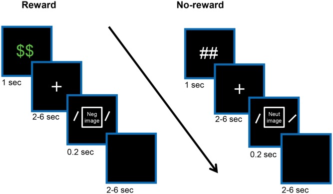
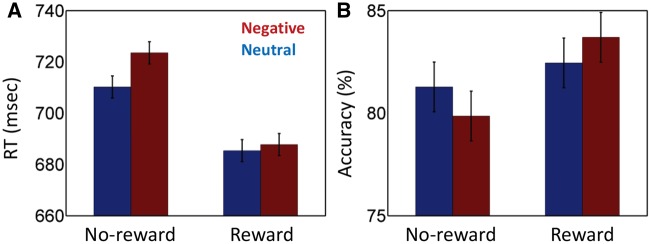
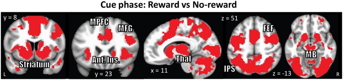
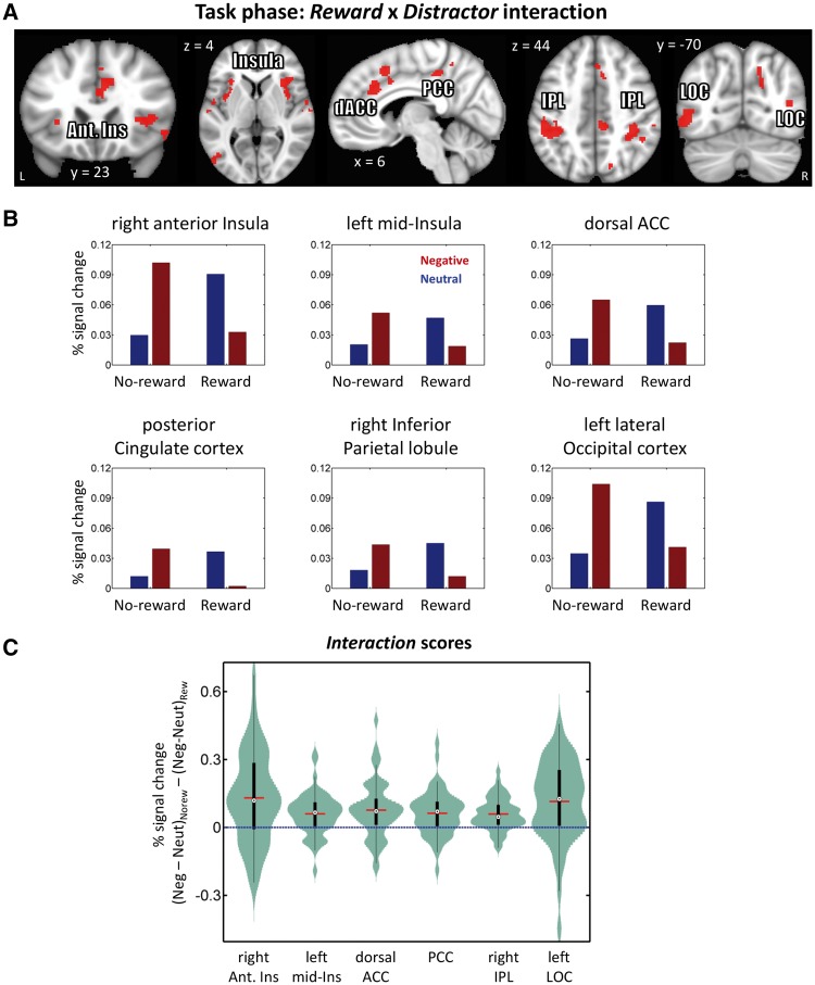
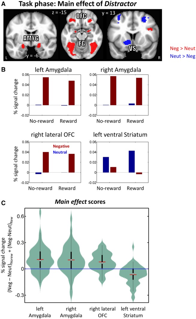
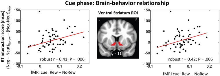
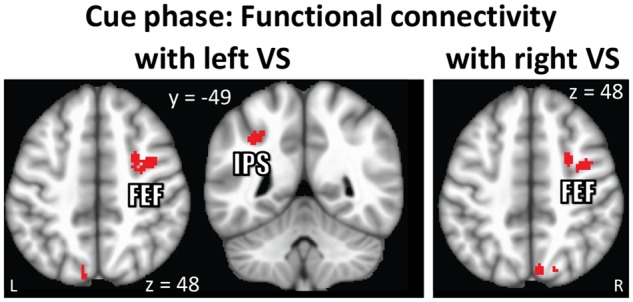
Knowledge about interactions between reward and negative processing is rudimentary. Here, we employed functional MRI to probe how potential reward signaled by advance cues alters aversive distractor processing during perception. Behaviorally, the influence of aversive stimuli on task performance was reduced during the reward compared to no-reward condition. In the brain, at the task phase, paralleling the observed behavioral pattern, we observed significant interactions in the anterior insula and dorsal anterior cingulate cortex, such that responses during the negative (vs neutral) condition were reduced during the reward compared to no-reward condition. Notably, negative distractor processing in the amygdala appeared to be independent of the reward manipulation. During the initial cue phase, we observed increased reward-related responses in the ventral striatum/accumbens, which were correlated with behavioral interference scores at the subsequent task phase, revealing that participants with increased reward-related responses exhibited a greater behavioral benefit of reward in reducing the adverse effect of negative images. Furthermore, during processing of reward (vs no-reward) cues, the ventral striatum exhibited stronger functional connectivity with fronto-parietal regions important for attentional control. Together, our findings contribute to the understanding of how potential reward influences attentional control and reduces negative distractor processing in the human brain.

Keywords: amygdala; emotion; perception; reward; ventral striatum.


Figures

Fig. 1.
Fig. 1.
Experimental design. On each trial, an initial cue indicated potential for reward followed by a variable delay period during which a white fixation cross was shown. Then a negative or neutral task-irrelevant picture (not shown here) was presented centrally and two bars were presented peripherally (not drawn to scale). The participant’s task was to indicate whether the bars are of same or different orientation while ignoring the central picture. Finally, each trial ended with a variable inter-trial interval. During the reward condition (left side), participants were rewarded if performance was both fast and accurate.
Fig. 2.
Fig. 2.
Simulated functional MRI responses and potential cue-task overlap. (A) To simulate transient cue responses, the cue was assumed to be presented for 1 s (as in the experiment), followed by a 4.5-s delay period (which varied between 2 and 6 s in the experiment), and a 0.2-s task phase (indicated by the timeline). The overlap between cue and task responses is small. (B) To simulate sustained cue responses, the cue was assumed to the presented for 5.5 s and followed immediately by a 0.2-s task phase (indicated by the timeline). The overlap between cue and task responses is quite substantial. Simulated responses were generated by employing a canonical gamma variate hemodynamic response function (Cohen, 1997).
Fig. 3.
Fig. 3.
Behavioral results. (A) RT data. During no-reward trials, negative images slowed responses relative to neutral ones. This interference effect was reduced during the reward condition. (B) Accuracy data. During no-reward trials, negative images reduced accuracy relative to neutral ones. This effect was reduced during the reward condition. Error bars denote standard within-subject error term for interaction effects (Loftus and Masson, 1994).
Fig. 4
Fig. 4
Cue phase functional MRI results displayed at an uncorrected P = 0.001 and 48-voxel cluster extent (cluster-level alpha of 0.05). Because cluster-extent based thresholding was used for multiple comparisons correction, voxels are displayed using a binary threshold. For further rationale about using binary maps in the context of cluster-based thresholding, see Woo et al, (2014). Increased responses were observed during processing of reward (vs no-reward) cue in subcortical regions and fronto-parietal regions. MPFC: medial prefrontal cortex, MFG: middle frontal gyrus, Ant. Ins: anterior insula, Thal: Thalamus, MB: midbrain, FEF: frontal eye-fields, IPS: intraparietal sulcus.
Fig. 5.
Fig. 5.
Task phase functional MRI results. (A) Clusters exhibiting significant Reward (reward, no-reward) × Distractor (neutral, negative) interactions displayed at an uncorrected P = 0.001 and 48-voxel cluster extent (cluster-level alpha of 0.05). Because cluster-extent based thresholding was used for multiple comparisons correction, voxels are displayed using a binary threshold. For further rationale about using binary maps in the context of cluster-based thresholding, see Woo et al. (2014). (B) Average responses within clusters that exhibited interaction effects. As some clusters spanned large sectors, for illustration purposes, clusters were selected at an uncorrected P =0.0005 and 34-voxel cluster extent (cluster-level alpha of 0.05). To avoid circularity, error bars are not plotted. (C) Distributions of within-subject interaction scores in units of % signal change. The box plots within the violins indicate the interquartile range (first quartile to third quartile), red lines show the mean values, and black dots inside white circles show median values. Ant. Ins: anterior insula, IPL: inferior parietal lobule, dACC: dorsal anterior cingulate cortex, PCC: posterior cingulate cortex, LOC: lateral occipital cortex.
Fig. 6.
Fig. 6.
Task phase functional MRI results displayed at an uncorrected P = 0.001 and 48-voxel cluster extent (cluster-level alpha of 0.05). Because cluster-extent based thresholding was used for multiple comparisons correction, voxels are displayed using a binary threshold. For further rationale about using binary maps in the context of cluster-based thresholding, see Woo et al. (2014). (A) Clusters exhibiting significant main effect of Distractor type (neutral, negative). (B) Main-effect response pattern from a subset of regions. To avoid circularity, error bars are not plotted. (C) Distributions of within-subject main effect of Distractor scores in units of % signal change. The box plots within the violins indicate the interquartile range (first quartile to third quartile), red lines show the mean values, and black dots inside white circles show median values. Amyg: amygdala, OFC: orbital frontal cortex, VS: ventral striatum, FG: fusiform gyrus.
Fig. 7.
Fig. 7.
Brain-behavior relationship. A positive linear relationship was observed between reward (vs no-reward) cue responses and behavioral RT interaction scores in the right and left ventral striatum ROIs.
Fig. 8.
Fig. 8.
Cue phase functional connectivity results displayed at an uncorrected P = 0.001 and 48-voxel cluster extent (cluster-level alpha of 0.05). Because cluster-extent based thresholding was used for multiple comparisons correction, voxels are displayed using a binary threshold. For further rationale about using binary maps in the context of cluster-based thresholding, see Woo et al. (2014). (A) Stronger functional coupling was observed between ventral striatum and fronto-parietal regions during the processing of reward relative to no-reward cues. VS: ventral striatum, FEF: frontal eye-fields, IPS: intraparietal sulcus.

References

    1. Bissonette G.B., Gentry R.N., Padmala S., Pessoa L., Roesch M.R. (2014). Impact of appetitive and aversive outcomes on brain responses: linking the animal and human literatures. Frontiers in Systems Neuroscience, 8, 24. - PMC - PubMed
    1. Boehler C.N., Hopf J.M., Stoppel C.M., Krebs R.M. (2012). Motivating inhibition: reward prospect speeds up response cancellation. Cognition, 125, 498–503. - PubMed
    1. Botvinick M., Braver T. (2015). Motivation and cognitive control: from behavior to neural mechanism. Psychology, 66, 83. - PubMed
    1. Braem S., Verguts T., Roggeman C., Notebaert W. (2012). Reward modulates adaptations to conflict. Cognition, 125, 324–32. - PubMed
    1. Chiew K.S., Braver T.S. (2013). Temporal dynamics of motivation-cognitive control interactions revealed by high-resolution pupillometry. Frontiers in Psychology, 4, 15. - PMC - PubMed