Review
. 2017 Jun:44:144-151.
doi: 10.1016/j.conb.2017.04.009. Epub 2017 May 12.

Sleep in the northern fur seal

Affiliations
Review

Sleep in the northern fur seal

Oleg I Lyamin et al. Curr Opin Neurobiol. 2017 Jun.

Abstract

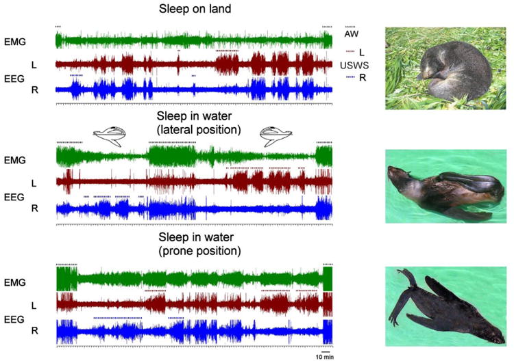
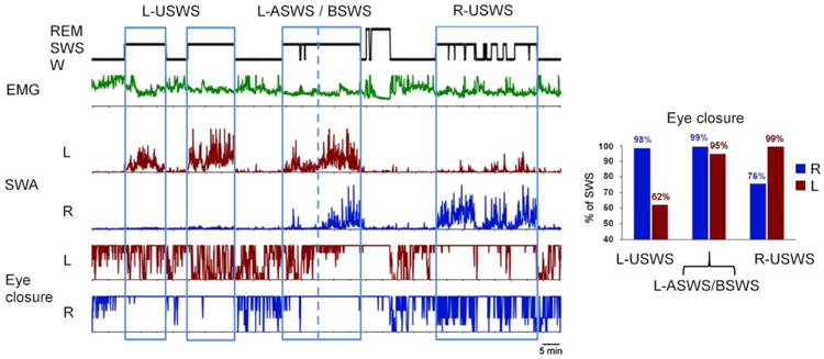
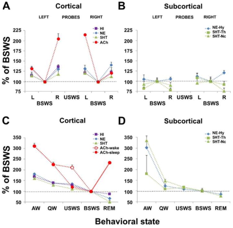
The pattern of sleep in the fur seal, a semiaquatic pinniped, has several striking behavioral and physiological adaptations that allow this species to inhabit both the land and water environment. These features include unihemispheric slow wave sleep (USWS, also being unihemispheric waking), the ability to maintain movement for stabilization of the sleep posture and to briefly open one eye while having a sleep electroencephalogram (EEG) in one hemisphere. In vivo microdialysis studies suggest that acetylcholine release is required for cortical activation during USWS, and that monoamines are not required for USWS. The need to breathe, to maintain efficient thermoregulation, and to avoid predation have shaped the sleep patterns in semiaquatic fur seals as in fully aquatic cetaceans.


Conflict of interest statement

Conflict of interest statement: We have no conflicts of interest.

Figures

Figure 1
Figure 1
Representative polygrams of recording from fur seals sleeping on land, at the surface in the lateral (middle) and in a prone position (bottom). The duration of each polygram is approximately 6 hr. EMG – neck electromyogram, EEG – electroencephalogram in symmetrical left (L) and (R) fronto-occipital derivations. Episodes of unihemispheric sleep (USWS) in the left and right hemispheres and active wakefulness (AW) are marked by brown, blue and black dotted lines, respectively. Postures of fur seals are shown on the photographs.
Figure 2
Figure 2
Sleep ontogeny in the fur seal. The diagrams show the amounts of sleep stages (REM sleep -rapid eye movement sleep, SWS - slow wave sleep, TST - total sleep time), duration and number of REM sleep episodes, and proportion/ ratio of asymmetrical SWS (ASWS) in fur seals of different ages (10-20 day old pups, 2-3 month old pups and in the group of juvenile males and females, 3-10 year old animals) while sleeping on land. The data are means + SEM calculated for 4, 4 and 7 individual fur seals from the 3 age groups.
Figure 3
Figure 3
Association between sleep and eye states in a fur seal. The diagrams on the left side (from top to bottom) show behavioral states (W- wakefulness, SWS – slow wave sleep and REM – rapid eye movement sleep), integrated neck electromyogram (EMG), slow wave activity (SWA, EEG power in the range of 1.2–4 Hz) in the left (L) and right (R) cerebral hemispheres and the time of closure of the left (L) and right (R) eyes. All parameters were scored or calculated for consecutive 5-sec epochs. Time scale is 5 min. Blue boxes highlight episodes of unihemispheric and asymmetrical SWS (USWS and ASWS) in the left and right hemispheres (L-USWS and R-USWS), and an episode of L-ASWS transitioned into bilateral SWS (BSWS). The blue dotted line separates the margin between L-ASWS and BSWS. The diagram on the right side shows percentage of time each eye was closed during the two episodes of L-USWS combined, episode of L-ASWS / BSWS and the episode of R-USWS as marked on the left diagram.
Figure 4
Figure 4
Release of neurotransmitters during sleep and waking states in the fur seal. (A,B) Cortical and subcortical release of histamine (HI), norepinephrine (NE), serotonin (5HT) and acetylcholine (ACh) during bilateral slow wave sleep (BSWS) and unihemispheric slow wave sleep (USWS) in the left (L) and right (R) hemispheres in fur seals. Each point represents the mean of the percent change in neurotransmitter level relative to BSWS which is calculated separately for the probes located in the left and right cortical hemispheres and subcortical structures (Hy, hypothalamus; Th, thalamus; Nc, caudate nucleus). (C,D) Cortical and subcortical release of HI, NE, 5HT, and ACh during waking and sleep states in fur seals. Each point represents the mean of the percent change in neurotransmitter level relative to BSWS. The release of HI, NE, and 5HT were not lateralized during USWS. It is shown as average for all left and right probes. The release of ACh was lateralized during USWS. For USWS it is shown separately for the waking (open circle) and sleeping (closed circle) hemispheres. During USWS, ACh release is higher on the waking side than on the sleeping side. The release of cortical HI, NE, and 5HT as well as subcortical NE and 5HT during USWS are not lateralized and are comparable to the release of ACh on the sleeping side. The data for cortical ACh and 5HT release were collected during the prior studies (reproduced from Lyamin et al., 2016).

Similar articles

Cited by

References

    1. Siegel JM. Do all animals sleep? Trends in Neurosciences. 2008;31:208–213. - PMC - PubMed
    1. Siegel JM. REM sleep: a biological and psychological paradox. Sleep Med Rev. 2011;15:139–42. - PMC - PubMed
    1. Siegel JM. Rapid eye movement sleep. In: Kryger MH, Roth T, Dement WC, editors. Principles and Practices of Sleep Mechanisms. Vol. 2016. WB Saunders; Philadelphia, PA: pp. 78–95.
    1. Joiner WJ. Unraveling the Evolutionary Determinants of Sleep. Curr Biol. 2016;26:R1073–R1087. - PMC - PubMed
    1. Heithaus MR, Frid A. Optimal diving under the risk of predation. J Theor Biol. 2003;223:79–92. - PubMed