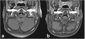
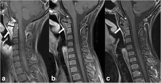
Clinical and MRI outcome of cervical spine lesions in children with juvenile idiopathic arthritis treated with anti-TNFα drugs early in disease course
- PMID: 28506237
- PMCID: PMC5433237
- DOI: 10.1186/s12969-017-0173-1
Clinical and MRI outcome of cervical spine lesions in children with juvenile idiopathic arthritis treated with anti-TNFα drugs early in disease course
Abstract
Backgrounds: The purpose of the study was to evaluate the clinical and magnetic resonance imaging (MRI) outcome of cervical spine arthritis in children with juvenile idiopathic arthritis (JIA), who received anti-TNFα early in the course of cervical spine arthritis.
Methods: Medical charts and imaging of JIA patients with cervical spine involvement were reviewed in this retrospective study. Data, including age at disease onset, JIA type, disease activity, treatment and clinical outcome were collected. Initial and followup MRI examinations of cervical spine were performed according to the hospital protocol to evaluate the presence of inflammation and potential chronic/late changes.
Results: Fifteen JIA patients with MRI proved cervical spine inflammation (11 girls, 4 boys, median age 6.3y) were included in the study: 9 had polyarthritis, 3 extended oligoarthritis, 2 persistent oligoarthritis and 1 juvenile psoriatic arthritis. All children were initially treated with high-dose steroids and methotrexate. In addition, 11 patients were treated with anti-TNFα drug within 3 months, and 3 patients within 7 months of cervical spine involvement confirmed by MRI. Mean observation time was 2.9y, mean duration of anti-TNFα treatment was 2.2y. Last MRI showed no active inflammation in 12/15 children, allowing to stop biological treatment in 3 patients, and in 3/15 significant reduction of inflammation. Mild chronic changes were found on MRI in 3 children.
Conclusions: Early treatment with anti-TNFα drugs resulted in significantly reduced inflammation or complete remission of cervical spine arthritis proved by MRI, and prevented the development of serious chronic/late changes. Repeated MRI examinations are suggested in the follow-up of JIA patients with cervical spine arthritis.
Keywords: Anti-TNFα; Cervical spine arthritis; Juvenile idiopathic arthritis; Magnetic resonance; Outcome.
Figures


References
-
- Wolfs JF, Arts MP, Peul WC. Juvenile chronic arthritis and the craniovertebral junction in the paediatric patient: review of the literature and management considerations. Adv Tech Stand Neurosurg. 2014;41:143–156. - PubMed
MeSH terms
Substances
LinkOut - more resources
Full Text Sources
Other Literature Sources
Medical

