. 2017 Jul 6;21(1):120-134.e7.
doi: 10.1016/j.stem.2017.03.024. Epub 2017 May 11.

Recruited Monocytes and Type 2 Immunity Promote Lung Regeneration following Pneumonectomy

Affiliations

Recruited Monocytes and Type 2 Immunity Promote Lung Regeneration following Pneumonectomy

Andrew J Lechner et al. Cell Stem Cell. .

Abstract

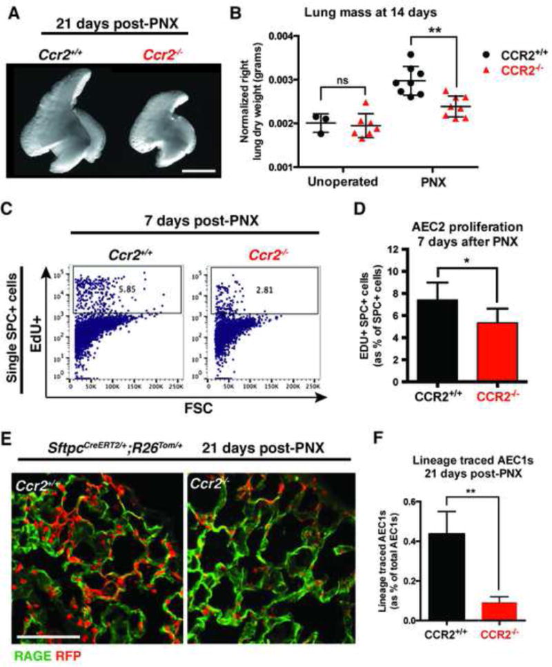
To investigate the role of immune cells in lung regeneration, we used a unilateral pneumonectomy model that promotes the formation of new alveoli in the remaining lobes. Immunofluorescence and single-cell RNA sequencing found CD115+ and CCR2+ monocytes and M2-like macrophages accumulating in the lung during the peak of type 2 alveolar epithelial stem cell (AEC2) proliferation. Genetic loss of function in mice and adoptive transfer studies revealed that bone marrow-derived macrophages (BMDMs) traffic to the lung through a CCL2-CCR2 chemokine axis and are required for optimal lung regeneration, along with Il4ra-expressing leukocytes. Our data suggest that these cells modulate AEC2 proliferation and differentiation. Finally, we provide evidence that group 2 innate lymphoid cells are a source of IL-13, which promotes lung regeneration. Together, our data highlight the potential for immunomodulatory therapies to stimulate alveologenesis in adults.

Keywords: ILC2; lung regeneration; macrophage; monocyte; pneumonectomy; type 2 alveolar pneumocyte.


Figures

Figure 1
Figure 1
Increased numbers of myeloid cells post-PNX. (A) Immunofluorescent staining of lung sections after PNX shows increased number of CSF1R-GFP+ cells (green, myeloid lineages) compared to unoperated animals. Scale bar = 100 um. (B) Flow cytometry shows increased numbers of CSF1R-GFP+,F4/80+ macrophages 7d post-PNX compared to sham operated animals. n=3 animals per group. Data shown are mean +/− SD. (C) Flow cytometry shows increased numbers of CD45+,,F4/80+,CD11b interstitial macrophages and monocytes 7d post-PNX compared to sham operated animals. (D) Flow cytometry shows increased numbers of CD45+,F4/80+,CD11c alveolar macrophages and monocytes 7d post-PNX compared to sham operated animals. (E, F) Quantification of data in C and D. n≥3 animals per group. Data shown are mean +/− SD. (G) Hierarchical clustering of 68 CD45+,CSF1R-GFP+,F4/80+,Ly6G-macrophages isolated from mouse lungs 7d post-PNX. At least 6 cell groups (x-axis) were defined by expression of 4 gene groups (y-axis). (H) Subpopulations of lung macrophages post-PNX included those which expressed high levels of monocyte markers (orange bar) and those which expressed high levels of M2-like macrophage markers (red bar). See also Figure S1.
Figure 2
Figure 2
CCR2+ monocytes are recruited to the lung post-PNX. (A) A cytokine protein array shows increased CCL2 7d post-PNX compared to sham operated animals. (B) Immunoflourescence shows increased numbers of Ccr2RFP+ monocytes 7d post-PNX compared to sham operated mice. (C) Flow cytometry shows increased numbers of CD45+,CD11b+,Ccr2FP+ cells 7d post-PNX compared to unoperated littermate controls. (D) Quantification of flow cytometry data shows increased numbers of CCR2+ monocytes after both PNX and sham operation compared to unoperated littermate controls. (E) Immunofluorescence on lungs from Ccr2RFP/RFP mice show decreased RFP+ monocytes 7d post-PNX compared to heterozygous mice (see B above). (F) Flow cytometry of dissociated lungs from Ccr2−/− mice shows decreased numbers of CD45+,F4/80+,CD11b+ interstitial monocytes and macrophages 7d post-PNX compared to wild type littermate controls. (G) Flow cytometry of dissociated lungs from Ccr2−/− mice shows that the number of CD45+,F4/80+,CD11c+ alveolar macrophages 7d post-PNX is not different from wild type littermate controls. For all experiments, n≥3 animals per group and data shown are mean +/− SD. Scale bars = 100 um. See also Figure S2.
Figure 3
Figure 3
CCR2+ monocytes are required for lung regeneration post-PNX. (A) Whole mount images of right accessory lobes 21d post-PNX show impaired regeneration in Ccr2−/− mice compared to wild type littermate controls. Scale bar = 5 mm. (B) Ccr2−/− mice show impaired lung regeneration assessed by dry weight of remaining right lobes 14d post-PNX compared to wild type littermates. (C, D) Flow cytometry shows decreased incorporation of EdU (24 h pulse) in AEC2 7d post-PNX in Ccr2−/− mice compared to wild type littermate controls. (E,F) Immunofluorescence on lung sections Sftpc-CreER;Rosa-tomato mice 21d post-PNX shows impaired differentiation of lineage labeled AEC2 (red) into AEC1 (RAGE, green) in Ccr2−/− mice compared to littermate controls. Scale bar = 100 um. For all experiments, n≥3 animals per group and data shown are mean +/− SD. See also Figure S3.
Figure 4
Figure 4
Macrophages support AEC2. (A) Ccr2−/− mice were given whole bone marrow from Ccr2+/+ or Ccr2−/− mice 4 and 7 d post-PNX by tail vein injection. Recipient mice were analyzed 14d post-PNX by lung dry weights. (B) Transfer of wild type bone marrow, but not bone marrow from Ccr2−/− mice, rescues the lung regeneration defect in CCR2-deficient mice. n≥3 mice per group, data shown are mean +/− SD. * = p ≤ 0.05; ** = p ≤ 0.01; *** = p ≤ 0.001 (C) Heat map and selected genes from RNA sequencing of pooled CD45+,F4/80+ macrophages isolated by FACS from wild type mice 7d post-PNX or sham operation. (D) Lineage labeled AEC2 were isolated by FACS from heterozygous Sftpc-CreER; Rosa-tomato mice and cultured for 14 days in 3-dimensions in varying ratios with primary CD45+,CSF1R-GFP+,F4/80+ macrophages from wild type mice; all wells contained 10,000 AEC2s, and either no macrophages (A), 5,000 macrophages (B), 10,000 macrophages (C), or 20,000 macrophages (D). (E, F) Macrophages have a dose-dependent effect on the colony formation of lineage labeled AEC2 in co-culture. N≥5 co-cultures for each ratio. Data shown are mean +/− SD. (G) Representative image of immunofluorescence on a histological section of a co-culture from condition D shows an F4/80+ macrophage in close association with a pneumosphere. Scale bar = 100 um. See also Figure S4.
Figure 5
Figure 5
IL4RA signaling is required for lung regeneration. (A) Immunofluorescent stains on sections from lungs of Arginase-YFP (YARG) mice show increased numbers of YARG+,F4/80+ M2-like macrophages (arrowheads) 4 and 7d post-PNX compared to sham. (B, C) Flow cytometry shows increased numbers of YARG+,F4/80+ M2-like macrophages 4 and 7d post-PNX compared to sham operated mice. (D–F) Immunofluorescence and flow cytometry on lungs from Il4ra−/− mice show rare YARG+; F4/80+ M2-like macrophages (arrowhead) 4d post-PNX compared to wild type YARG littermate controls (see A above). (G) Il4ra−/− mice show impaired lung regeneration assessed by dry weight of the right accessory lobe 14d post-PNX. ** = p ≤ 0.01. (H) Immunoflourescence on sections of lungs from Ccr2RFP/+;YARG mice show YARG+ M2-like macrophages that are both CCR2-RFP+ (arrows) and CCR2-RFP-(arrowheads). (I) Quantification of flow cytometry on dissociated lungs from Ccr2RFP/RFP; YARG mice show decreased numbers of YARG+, F4/80+ M2-like macrophages 4d post-PNX compared to wild type YARG controls. (J) CD45.1 wild type mice were lethally irradiated and given bone marrow from either wild type or Il4ra−/− (CD45.2) mice by tail vein injection. 10 weeks were allowed for hematopoietic reconstitution. PNX was performed on chimeras and they were analyzed by lung dry weight 14d later. (K) Il4ra−/− bone marrow recipients show impaired lung regeneration compared to recipients of wild type bone marrow. * = p ≤ 0.05. For all experiments, n≥3 animals per group and data shown are mean +/− SD. Scale bars = 100 um. See also Figure S5.
Figure 6
Figure 6
ILC2s accumulate and produce IL-13 post-PNX. (A) Immunofluorescent stains on sections from lungs of IL5RFP/+ (Red5) mice show ILC2s near small airways and in alveolar spaces post-PNX. Scale bar = 100 um. (B, C) Flow cytometry of dissociated lungs from Il5RFP/+ mice shows increased numbers of CD45+LinThy1+IL5+ ILC2s 7d post-PNX compared to sham operated animals. (D, E) Flow cytometry of dissociated lungs from Smart13 mice shows increased numbers of IL-13 producing ILC2s 4d post-PNX compared to sham operated animals. n≥3 animals for each group. Data are represented as mean +/− SD. (F) Proposed model demonstrating the roles of CCR2+ monocytes, M2-like macrophages, and ILC2s in PNX-induced lung regeneration. See also Figure S6.

Comment in

References

    1. Aggarwal NR, King LS, D’Alessio FR. Diverse macrophage populations mediate acute lung inflammation and resolution. Am J Physiol Lung Cell Mol Physiol. 2014;306:L709–25. - PMC - PubMed
    1. Anders S, Pyl PT, Huber W. HTSeq–a Python framework to work with high-throughput sequencing data. Bioinformatics. 2015;31:166–169. - PMC - PubMed
    1. Arnold L, Henry A, Poron F, Baba-Amer Y, van Rooijen N, Plonquet A, Gherardi RK, Chazaud B. Inflammatory monocytes recruited after skeletal muscle injury switch into antiinflammatory macrophages to support myogenesis. J Exp Med. 2007;204:1057–1069. - PMC - PubMed
    1. Aurora A, Olson E. Immune Modulation of Stem Cells and Regeneration. Cell Stem Cell. 2014;15:1425. - PMC - PubMed
    1. Aurora AB, Porrello ER, Tan W, Mahmoud AI, Hill JA, Bassel-Duby R, Sadek HA, Olson EN. Macrophages are required for neonatal heart regeneration. J Clin Invest. 2014;124:1382–1392. - PMC - PubMed

Publication types

MeSH terms