. 2017 Jun 6;8(6):1506-1515.
doi: 10.1016/j.stemcr.2017.04.013. Epub 2017 May 11.

NEUROD1 Instructs Neuronal Conversion in Non-Reactive Astrocytes

Affiliations

NEUROD1 Instructs Neuronal Conversion in Non-Reactive Astrocytes

Rebecca Brulet et al. Stem Cell Reports. .

Abstract

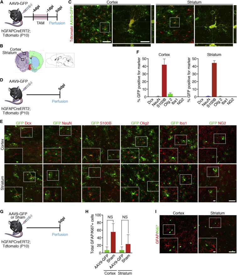
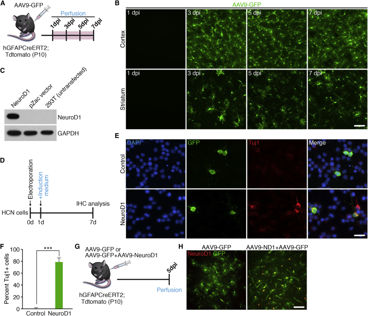
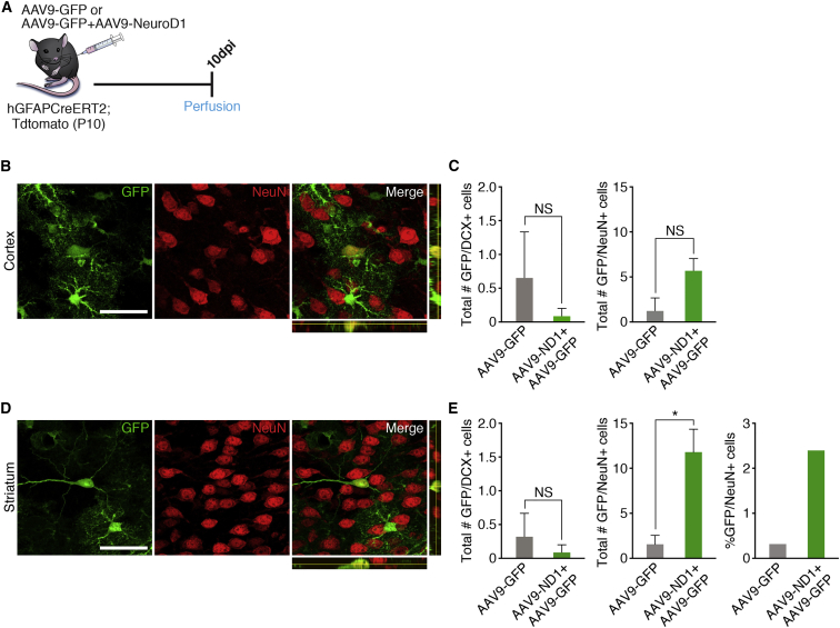
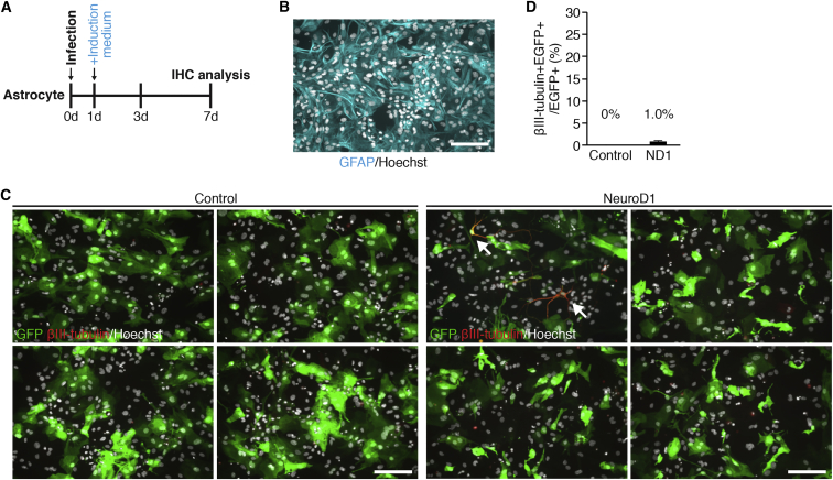
Currently, all methods for converting non-neuronal cells into neurons involve injury to the brain; however, whether neuronal transdifferentiation can occur long after the period of insult remains largely unknown. Here, we use the transcription factor NEUROD1, previously shown to convert reactive glial cells to neurons in the cortex, to determine whether astrocyte-to-neuron transdifferentiation can occur under physiological conditions. We utilized adeno-associated virus 9 (AAV9), which crosses the blood-brain barrier without injury, to deliver NEUROD1 to astrocytes through an intravascular route. Interestingly, we found that a small, but significant number of non-reactive astrocytes converted to neurons in the striatum, but not the cortex. Moreover, astrocytes cultured to minimize their proliferative potential also exhibited limited neuronal transdifferentiation with NEUROD1 expression. Our results show that a single transcription factor can induce astrocyte-to-neuron conversion under physiological conditions, potentially facilitating future clinical approaches long after the acute injury phase.

Keywords: AAV9; NeuroD; astrocytes; cortex; neurons; reprogramming; striatum; transdifferentiation.


Figures

Figure 1
Figure 1
AAV9-GFP Labels Neocortical and Striatal Astrocytes in Postnatal Day 10 Mouse Brain (A) Timeline showing the experimental design. (B) Schematic of the brain showing the areas in which representative images were taken. (C) Representative images of AAV9-GFP infection in the cortex and striatum at 5 days post injection (dpi). (D) Timeline showing experimental design for validating identity of GFP+ cells in (E). (E) Representative images showing co-labeling of GFP+ cells with the neuronal markers DCX and NeuN, the astrocyte marker S100b, the oligodendrocyte precursor marker OLIG2, the microglial marker IbaI, and the NG2 glial marker NG2. (F) Quantification of overlap of GFP+ cells for respective markers in the cortex and striatum. n = 3 mice/marker. Data are shown as mean ± SEM. (G) Timeline showing the experimental design. (H) Quantification of GFAP/Ki67+ reactive astrocytes in the cortex and striatum 5 dpi. n = 3 mice/group. Data are shown as mean ± SEM. NS, not significant. (I) Representative images showing sections of the cortex and striatum stained for GFAP and Ki67. Scale bars represent 50 μm in main panels and 25 μm in insets.
Figure 2
Figure 2
Confirmation of NEUROD1 Overexpression in Neocortical and Striatal Astrocytes (A) Timeline showing experimental design. (B) Representative images taken from the cortex and striatum showing AAV9-GFP infection from 1 to 7 dpi. Scale bar, 50 μm. (C) Western blot showing successful overexpression of NEUROD1 after cloning into the AAV9 backbone. (D) Timeline showing experimental design. (E) Representative in vitro images of electroporated cells, 6 days post electroporation, with either AAV9-ND1 + pllU2g-GFP or pllU2g-GFP alone. Scale bar, 50 μm. (F) Quantification of GFP/Tuj1+ cells at 6 days post electroporation. ∗∗∗p = 0.0002. n = 3 independent experiments. (G) Timeline showing experimental design. (H) Representative images from the cortex showing significant overlap of infected astrocytes and NEUROD1 in mice injected with AAV9-NEUROD1, but not AAV9-GFP control, at 5 dpi. Scale bar, 50 μm.
Figure 3
Figure 3
NEUROD1 Expression in Striatal Astrocytes Led to Limited Neuronal Conversion (A) Timeline showing experimental design. Mice at 10 days old were injected with either AAV9-NEUROD1 + AAV9-GFP (experimental) or AAV9-GFP (control) and euthanized at 10 dpi to determine whether neuronal conversion had occurred. (B) Representative images showing one of the few cells expressing NeuN in the cortex. Scale bar, 50 μm. (C) Quantification showing the total number of GFP+ cells that co-labeled for the neuronal markers DCX and NeuN in the cortex. n = 3 mice for AAV9-GFP, n = 11 mice for AAV9-NEUROD1 + AAV9-GFP. Data are shown as mean ± SEM. NS, not significant. (D) Representative images showing one of the few cells expressing NeuN in the striatum. Scale bar, 50 μm. (E) Quantification showing the total number and percentage of GFP+ cells that co-labeled with the neuronal markers DCX and NeuN in the striatum. n = 3 mice for AAV9-GFP, n = 11 mice for AAV9-NEUROD1 + AAV9-GFP. Data are shown as mean ± SEM. p < 0.05; NS, not significant.
Figure 4
Figure 4
Overexpression of NEUROD1 Resulted in Minimal Neuronal Differentiation In Vitro (A) Timeline showing experimental design. (B) Representative image of cultured astrocytes showing expression of the astrocyte marker GFAP. Scale bar, 50 μm. (C) Representative images taken in vitro showing astrocytes infected with lenti-CAG-NEUROD1-UbiC-GFP or lenti-CAG-UbiC-GFP at 7 dpi co-labeled with the immature neuronal marker βIII-tubulin (Tuj1). Arrows denote GFP+/Tuj1+ cells. Scale bar, 50 μm. (D) Graph showing the percentage of all astrocytes expressing Tuj1 7 days after infection. n = 4 independent experiments/condition. Data are expressed as mean ± SEM.

References

    1. Anderson M.A., Ao Y., Sofroniew M.V. Heterogeneity of reactive astrocytes. Neurosci. Lett. 2014;565:23–29. - PMC - PubMed
    1. Aravantinou-Fatorou K., Ortega F., Chroni-Tzartou D., Antoniou N., Poulopoulou C., Politis P.K., Berninger B., Matsas R., Thomaidou D. CEND1 and NEUROGENIN2 reprogram mouse astrocytes and embryonic fibroblasts to induced neural precursors and differentiated neurons. Stem Cell Reports. 2015;5:405–418. - PMC - PubMed
    1. Barreto G.E., Sun X., Xu L., Giffard R.G. Astrocyte proliferation following stroke in the mouse depends on the distance from the infarct. PLoS One. 2011;6:e27881. - PMC - PubMed
    1. Bayraktar O.A., Fuentealba L.C., Alvarez-Buylla A., Rowitch D.H. Astrocyte development and heterogeneity. Cold Spring Harb. Perspect. Biol. 2015;7:1–16. - PMC - PubMed
    1. Buffo A., Inmaculada R., Tripathi P., Lepier A., Colak D., Horn A., Mori T., Götz M. Origin and progeny of reactive gliosis: a source of multipotent cells in the injured brain. Proc. Natl. Acad. Sci. USA. 2008;105:3581–3586. - PMC - PubMed

MeSH terms

Substances