

Hepatic stellate cells as key target in liver fibrosis
- PMID: 28506744
- PMCID: PMC5682243
- DOI: 10.1016/j.addr.2017.05.007
Hepatic stellate cells as key target in liver fibrosis
Abstract
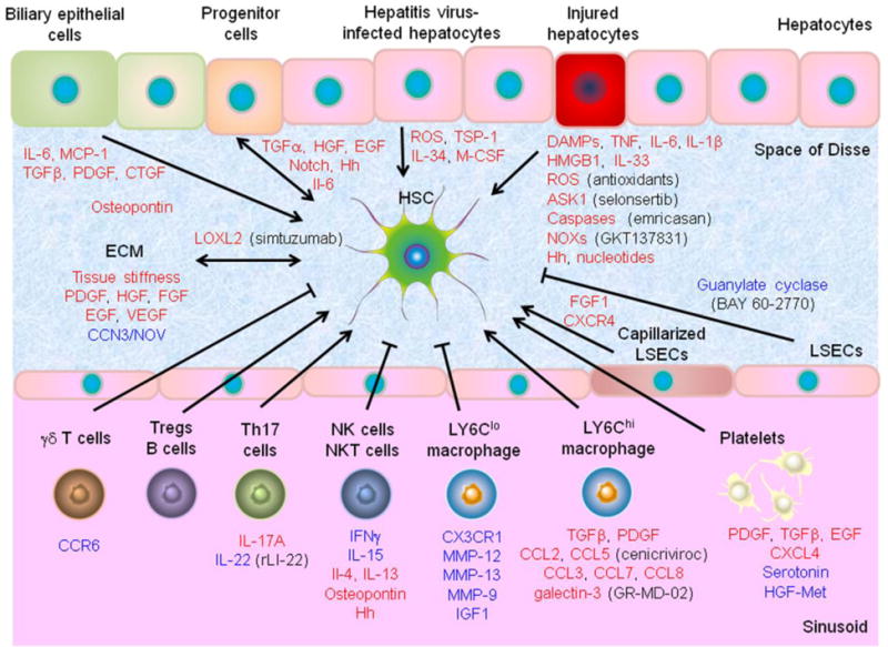
Progressive liver fibrosis, induced by chronic viral and metabolic disorders, leads to more than one million deaths annually via development of cirrhosis, although no antifibrotic therapy has been approved to date. Transdifferentiation (or "activation") of hepatic stellate cells is the major cellular source of matrix protein-secreting myofibroblasts, the major driver of liver fibrogenesis. Paracrine signals from injured epithelial cells, fibrotic tissue microenvironment, immune and systemic metabolic dysregulation, enteric dysbiosis, and hepatitis viral products can directly or indirectly induce stellate cell activation. Dysregulated intracellular signaling, epigenetic changes, and cellular stress response represent candidate targets to deactivate stellate cells by inducing reversion to inactivated state, cellular senescence, apoptosis, and/or clearance by immune cells. Cell type- and target-specific pharmacological intervention to therapeutically induce the deactivation will enable more effective and less toxic precision antifibrotic therapies.
Keywords: Alcoholic liver disease; Cirrhosis; Hepatitis; Myofibroblast; Non-alcoholic fatty liver disease; Non-alcoholic steatohepatitis.
Copyright © 2017 Elsevier B.V. All rights reserved.
Figures

References
-
- Rockey DC, Bell PD, Hill JA. Fibrosis–a common pathway to organ injury and failure. N Engl J Med. 2015;372:1138–1149. - PubMed
-
- G.B.D.R.F. Collaborators. Forouzanfar MH, Alexander L, Anderson HR, Bachman VF, Biryukov S, et al. Global, regional, and national comparative risk assessment of 79 behavioural, environmental and occupational, and metabolic risks or clusters of risks in 188 countries, 1990–2013: a systematic analysis for the Global Burden of Disease Study 2013. Lancet. 2015;386:2287–2323. - PMC - PubMed
-
- Tsochatzis EA, Bosch J, Burroughs AK. Liver cirrhosis. Lancet. 2014;383:1749–1761. - PubMed
-
- Ge PS, Runyon BA. Treatment of Patients with Cirrhosis. N Engl J Med. 2016;375:767–777. - PubMed
Publication types
MeSH terms
Grants and funding
LinkOut - more resources
Full Text Sources
Other Literature Sources
Medical
