. 2017 May 30;114(22):E4425-E4434.
doi: 10.1073/pnas.1702909114. Epub 2017 May 15.

Spatially restricted dental regeneration drives pufferfish beak development

Affiliations

Spatially restricted dental regeneration drives pufferfish beak development

Alexandre P Thiery et al. Proc Natl Acad Sci U S A. .

Abstract

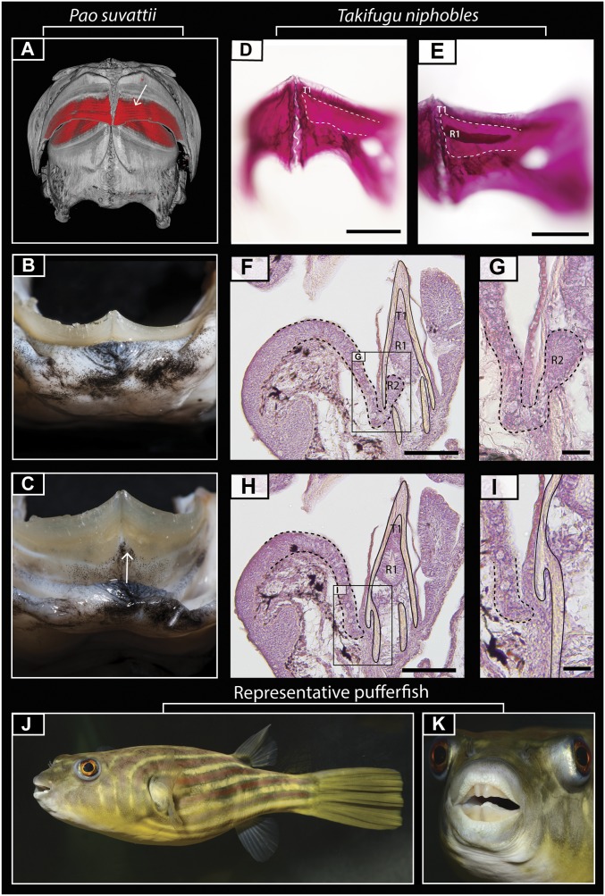
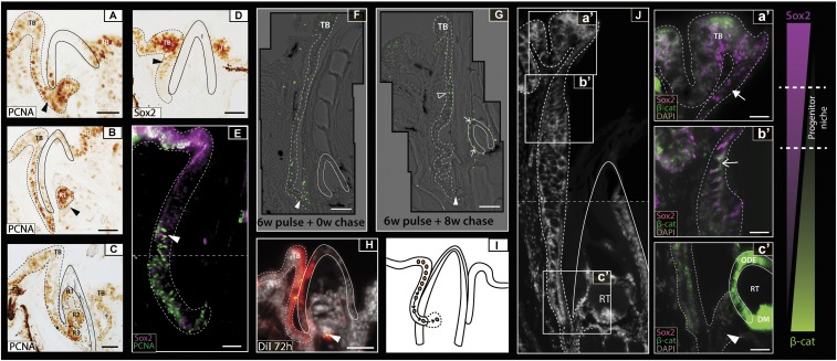
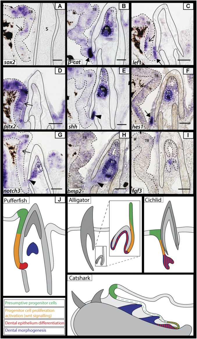
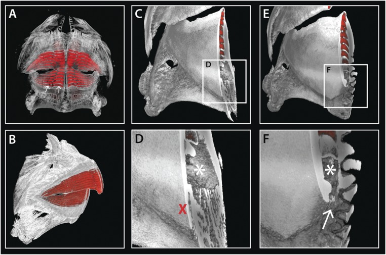
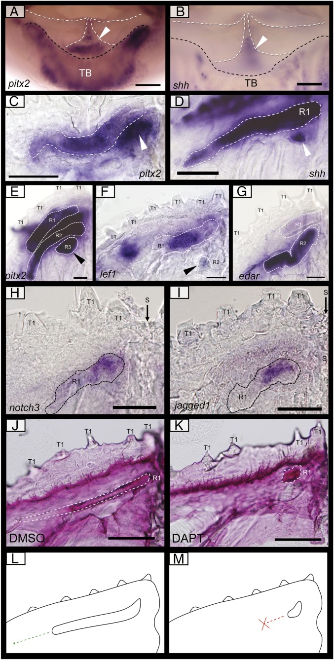
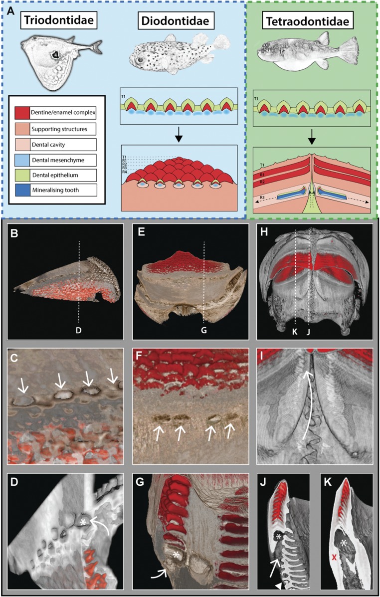
Vertebrate dentitions are extraordinarily diverse in both morphology and regenerative capacity. The teleost order Tetraodontiformes exhibits an exceptional array of novel dental morphologies, epitomized by constrained beak-like dentitions in several families, i.e., porcupinefishes, three-toothed pufferfishes, ocean sunfishes, and pufferfishes. Modification of tooth replacement within these groups leads to the progressive accumulation of tooth generations, underlying the structure of their beaks. We focus on the dentition of the pufferfish (Tetraodontidae) because of its distinct dental morphology. This complex dentition develops as a result of (i) a reduction in the number of tooth positions from seven to one per quadrant during the transition from first to second tooth generations and (ii) a dramatic shift in tooth morphogenesis following the development of the first-generation teeth, leading to the elongation of dental units along the jaw. Gene expression and 1,1'-Dioctadecyl-3,3,3',3'-tetramethylindocarbocyanine perchlorate (DiI) lineage tracing reveal a putative dental epithelial progenitor niche, suggesting a highly conserved mechanism for tooth regeneration despite the development of a unique dentition. MicroCT analysis reveals restricted labial openings in the beak, through which the dental epithelium (lamina) invades the cavity of the highly mineralized beak. Reduction in the number of replacement tooth positions coincides with the development of only four labial openings in the pufferfish beak, restricting connection of the oral epithelium to the dental cavity. Our data suggest the spatial restriction of dental regeneration, coupled with the unique extension of the replacement dental units throughout the jaw, are primary contributors to the evolution and development of this unique beak-like dentition.

Keywords: dental regeneration; diversity; novelty; stem cells; tooth development.


Conflict of interest statement

The authors declare no conflict of interest.

Figures

Fig. 1.
Fig. 1.
Pufferfish dental morphology. (A) Reconstructed microCT scan of adult P. suvattii reveals a banded dentition encased in osteodentine. (B and C) Digital photos show the fleshy lip of P. suvattii and its attachment to the beaked dentition at the fused beak/dentary boundary. A cleft can be seen at the symphysis of the jaw, with the attachment of the labial oral epithelium to the beak following the contour of the cleft (white arrow in C). (D and E) Alizarin red staining of early embryonic T. niphobles samples reveals a single developing band in each jaw quadrant. (FI) Hematoxylin-stained sagittal paraffin sections reveal a gubernacular opening at the osteodentine/mandibular boundary in each jaw quadrant (F and G) (depicted by dashed lines in H) that is absent in serial sections of the adjacent areas (H and I). G and I are close-up images of the boxed regions in F and H, respectively. In the histological sections, a continuous stream of epithelium connects the oral epithelium to the regenerating teeth (R1 and R2) within the dental cavity. (J and K) Digital photographs of Tetraodon lineatus showing the overall morphology of a “typical” pufferfish. R1-2, replacement tooth generations; T1, first tooth generation. (Scale bars: 250 µm in D and E; 100 µm in F and H; 50 µm in G and I.)
Fig. 2.
Fig. 2.
Localization of a dental progenitor cell niche within the pufferfish dental lamina. (AC) P. baileyi PCNA immunohistochemistry reveals high levels of cellular proliferation within the oral epithelium. As replacement teeth progress from late-initiation (A) to morphogenesis (C), the new tooth generation (R1) buds from the dental lamina (B). Successive rounds of replacement show that the dental generations stack on one another within an enameloid outer casing (black line) (C). (D) Sox2 immunohistochemical labeling during dental replacement initiation depicts high levels of Sox2 within both the developing taste buds (TB) and the dental progenitor site located within the labial oral epithelium (dental lamina) (black arrowhead). (E) Double immunofluorescence treatment for Sox2/PCNA in T. niphobles shows low levels of PCNA expression within the Sox2+ cells of the presumptive dental progenitor niche and cells within the aboral dental lamina exhibiting high levels of PCNA. The horizontal dashed line depicts image stitching of two adjacent images. White arrowhead marks region of overlapping PCNA/Sox2 expression. (F) BrdU pulse/chase experiments (0.2 mM) show the incorporation of BrdU into dividing cells after 6 wk of treatment, with high levels of incorporation noted in the distal dental lamina next to the base of the beak (white arrowhead). (G) After a further 8-wk chase, label-retaining cells were found in the most superficial dental lamina cells (open arrowhead) but not in the distal dental lamina (white arrowhead). Label-retaining cells found in the dental epithelium of the developing tooth are indicated by a white arrow. Images in F and G are composites of multiple images taken at high magnification and stitched together. (H) DiI labeling of the labial oral epithelium in P. suvattii highlighted this region as a presumptive source of dental progenitor cells. DiI was detected within the outer dental epithelium of the tooth (white arrowhead) 72 h post DiI treatment. (I) As summarized in a schematic representation, we observed a continuous field of Sox2+ cells between the labial taste bud and the dental progenitor site, with cells from the latter migrating and contributing to the new dental generations. Black arrows represent the direction of cell movement. (J) Sox2/ABC double immunohistochemical labeling on adult C. travancoricus highlights epithelial Sox2+/ABC (a′, white filled arrow), Sox2+/ABC+ (b′, white arrow), and Sox2/ABC+ (c′, white arrowhead) regions within the dental lamina. Coexpression of these markers marks the site of activation of putative dental progenitors within the oral epithelium. Dashed line across (J) depicts image stitching of two adjacent images. Images are orientated with labial to the left and oral to the top. The dotted line in all images depicts the boundary of the oral epithelium and the end of the dental lamina. DM, dental mesenchyme; ODE, outer dental epithelium; R1–3, replacement tooth generations; RT, regenerating tooth; TB, labial taste bud. (Scale bars: 25 µm in AE; 20 µm in F, a′F, c′; 50 µm in G and H; 15 µm in I.)
Fig. 3.
Fig. 3.
Conserved odontogenic signaling regulates dental regeneration in pufferfish. (AI) Expression of well-documented odontogenic markers belonging to Sox (sox2, A); canonical Wnt signaling (β-catenin, B; lef1 C); Pitx (pitx2, D); Shh (E), Notch (hes1, F; notch3 G); Bmp (bmp2, H); and Fgf (fgf3, I) gene families in T. niphobles embryos. The thin arrow marks the site of presumptive dental progenitors, with expression of pitx2 (D), lef1 (C), and sox2 (A) within this region. The thick arrow marks the distal end of the dental lamina. The filled arrowhead highlights an opening within the osteodentine beak casing through which new odontogenic cells bud from the dental lamina. β-cat (B), shh (E), hes1 (F), notch3 (G), bmp2 (H), and fgf3 (I) are all expressed within the epithelium of the latest developing teeth. (J) A diagrammatic illustration of odontogenetically similar structures in various polyphyodonts [pufferfish, alligator (7, 16), cichlid (5), and catshark (43)]. Four main developmental regions are highlighted: presumptive dental progenitors, progenitor cell activation marked by the coexpression of Sox and Wnt signals, dental epithelium differentiation marked by the up-regulation of various developmental genes at the distal tip of the dental lamina and the growth of a tooth bud, and dental morphogenesis. The dotted line depicts the boundary of the oral epithelium and the end of the dental lamina. All images were taken from 14-µm sagittal paraffin-embedded sections. A, B, E, F, H, and I are from T. niphobles embryos at 50 dpf. C, D, and G are from embryos at 32 dpf. R1-2, replacement tooth generations; S, suture; TB, labial taste bud. (Scale bars: 50 µm in BD, F, and G; 35 µm in A, E, H, and I.)
Fig. S1.
Fig. S1.
Takifugu dental morphology. (A and B) Reconstructed microCT scans of adult T. niphobles reveal discontinuous dentine bands (white arrows in A) encased in osteodentine. (CF) Virtual slices through the dentition at both symphyseal (C and D) and lateral (E and F) regions reveal a single gubernacular opening (white arrow in F) in each jaw quadrant. An asterisk in D and F marks the dental cavity; red cross in D marks the absence of a gubernacular opening.
Fig. 4.
Fig. 4.
Gene-expression patterns during dental regeneration morphogenesis and chemical inhibition of Notch signaling through small-molecule treatments. Whole-mount RNA in situ hybridization of the lower jaws of P. baileyi at 57 dpf (A and B and E and F), 53 dpf (H and I), 46 dpf (G), and T. niphobles at 50 dpf (C and D), and P. suvattii at 26 dpf (J and K). (A and B) Images taken from above the lower jaw depict expression of pitx2 (A) and shh (B) in the labial epithelium at the jaw symphysis (white arrowhead). (CI) Images of a single lower-jaw quadrant, with the expression of pitx2 (C and E), shh (D), lef1 (F), edar (G), notch3 (H), and jagged1b (I) in the developing teeth illustrated through dotted lines (CG). New tooth units can be seen initially developing (CF, arrowhead) at the symphysis of the beak (right of image). (J and K) Alizarin red staining of P. suvattii embryos after treatment with 50 μM DAPT during initiation of the second-generation dentition for 72 h, followed by a 2-wk recovery period (K), and control sample treated with 1% DMSO (K). Staining reveals the mineralization of the second-generation tooth (R1, white dotted line), with the control specimens (J) elongating laterally throughout the jaw (n = 5/5). DAPT treatment (25 μM) (K) resulted in the loss of dental elongation, with the mineralized tooth restricted in size at its site of initiation (n = 7/7). (L and M) Schematic representation of the phenotypes observed in the DMSO and DAPT treatments described above. R1–3, replacement tooth generations; S, suture; T1, first tooth generation. (Scale bars: 100 µm in AF. 50 µm in GK.)
Fig. 5.
Fig. 5.
(A) Schematic highlighting the modes of dental replacement in Triodontidae, Diodontidae, and Tetraodontidae. Although the development of the first-generation teeth is conserved, the modes of dental replacement differ dramatically. R1–R4, replacement tooth generations; T1, first dental generation. Reconstructed microCT scans of Triodontidae (BD) and Diodontidae (EG) reveal gubernacular openings (C and F, white arrows) within the labial surface of the jawbone. Virtual sagittal sections through these sites show an open connection (white arrow) between the labial surface and the dental cavity (marked by an asterisk) (D and G). In Triodontidae and Diodontidae (BG), gubernacular openings can be seen associated with multiple tooth sites along the jaw. MicroCT scans of P. suvattii (H and I) illustrate an intraosseous, banded dentition, with virtual serial slices through the dentition at the parasymphyseal region (J) and adjacent region (K) revealing gubernacular openings restricted to the parasymphyseal region (white arrow in I and J, red cross in K), with a single opening in each jaw quadrant. These openings connect the site of dental lamina attachment (white arrow in I) with the dental cavity (marked by an asterisk). The white arrowhead in J marks the interdigitating jaw suture observable at the symphysis.

Similar articles

Cited by

References

    1. Wagner GP, Lynch VJ. Evolutionary novelties. Curr Biol. 2010;20:R48–R52. - PubMed
    1. Jernvall J, Thesleff I. Tooth shape formation and tooth renewal: Evolving with the same signals. Development. 2012;139:3487–3497. - PubMed
    1. Sire JY, Davit-Beal T, Delgado S, Van Der Heyden C, Huysseune A. First-generation teeth in nonmammalian lineages: Evidence for a conserved ancestral character? Microsc Res Tech. 2002;59:408–434. - PubMed
    1. Tucker AS, Fraser GJ. Evolution and developmental diversity of tooth regeneration. Semin Cell Dev Biol. 2014;25-26:71–80. - PubMed
    1. Fraser GJ, Bloomquist RF, Streelman JT. Common developmental pathways link tooth shape to regeneration. Dev Biol. 2013;377:399–414. - PMC - PubMed

Publication types

LinkOut - more resources