. 2017 May;5(9):e13281.
doi: 10.14814/phy2.13281. Epub 2017 May 14.

Matrix stiffness regulates migration of human lung fibroblasts

Affiliations

Matrix stiffness regulates migration of human lung fibroblasts

Shuichi Asano et al. Physiol Rep. 2017 May.

Abstract

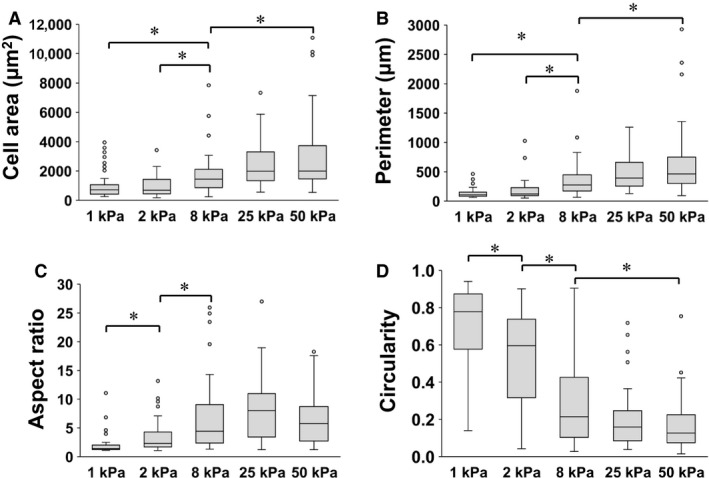
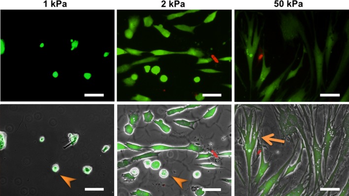
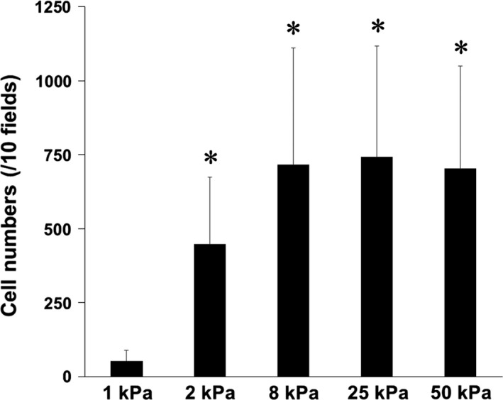
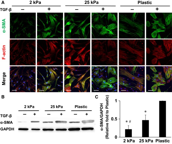
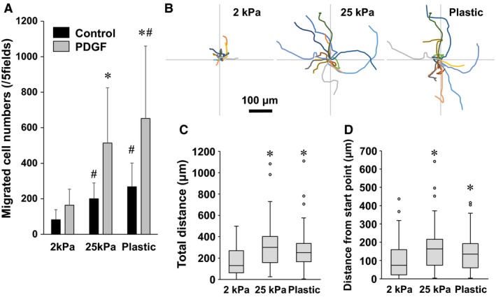
In patients with pulmonary diseases such as idiopathic pulmonary fibrosis and severe acute respiratory distress syndrome, progressive pulmonary fibrosis is caused by dysregulated wound healing via activation of fibroblasts after lung inflammation or severe damage. Migration of fibroblasts toward the fibrotic lesions plays an important role in pulmonary fibrosis. Fibrotic tissue in the lung is much stiffer than normal lung tissue. Emerging evidence supports the hypothesis that the stiffness of the matrix is not only a consequence of fibrosis, but also can induce fibroblast activation. Nevertheless, the effects of substrate rigidity on migration of lung fibroblasts have not been fully elucidated. We evaluated the effects of substrate stiffness on the morphology, α-smooth muscle actin (α-SMA) expression, and cell migration of primary human lung fibroblasts by using polyacrylamide hydrogels with stiffnesses ranging from 1 to 50 kPa. Cell motility was assessed by platelet-derived growth factor (PDGF)-induced chemotaxis and random walk migration assays. As the stiffness of substrates increased, fibroblasts became spindle-shaped and spread. Expression of α-SMA proteins was higher on the stiffer substrates (25 kPa gel and plastic dishes) than on the soft 2 kPa gel. Both PDGF-induced chemotaxis and random walk migration of fibroblasts precultured on stiff substrates (25 kPa gel and plastic dishes) were significantly higher than those of cells precultured on 2 kPa gel. Transfection of the fibroblasts with short interfering RNA for α-SMA inhibited cell migration. These findings suggest that fibroblast activation induced by a stiff matrix is involved in mechanisms of the pathophysiology of pulmonary fibrosis.

Keywords: Matrix stiffness; mechanotransduction; migration; pulmonary fibrosis; α‐smooth muscle actin.


Figures

Figure 1
Figure 1
Time‐dependent changes of cell morphology on different substrates. Representative phase‐contrast images of normal human lung fibroblasts cultured on polyacrylamide hydrogels of different stiffnesses (1, 2, and 50 kPa) for 4, 16, and 72 h. Cell growth and spreading were suppressed on the softest (1 kPa) gels. Arrowhead and arrow show nonspreading and spreading cells, respectively. Bar = 100 μm.
Figure 2
Figure 2
Effects of substrate stiffness on morphology of fibroblasts. Fibroblasts were cultured on polyacrylamide hydrogels of different stiffnesses (1, 2, 8, 25, and 50 kPa) for 16 h, and 50 cells each from three independent experiments were evaluated using ImageJ software. Cell area (A), perimeter (B), aspect ratio (C), and circularity (D) of the cells cultured on different stiffnesses (1, 2, 8, 25, and 50 kPa) of polyacrylamide hydrogels are compared. Boxes represent the 25th and 75th percentiles; whiskers indicate 10th and 90th percentiles. *Significantly different between the groups (P < 0.05).
Figure 3
Figure 3
Effects of substrate stiffness on cell viability. Upper images: Representative fluorescent images of fibroblasts cultured on 1 (left), 2 (middle), and 50 kPa (right) polyacrylamide hydrogels for 2 days. Lower images: Merged images of the fluorescent and phase‐contrast images. Live cells were stained with fluorescent calcein (green) and dead cells with ethidium homodimer‐1 (red). Nonspreading cells on the 1 and 2 kPa gels (arrows in left and middle images) were still alive. The arrowhead (right image) indicates a spread cell. Bar = 100 μm.
Figure 4
Figure 4
Effects of substrate stiffness on cell proliferation. Lung fibroblasts were cultured on different stiffness of polyacrylamide hydrogels for 72 h. The numbers of cells per 10 fields were manually counted. Values are means ± SD of six independent experiments. *< 0.05 vs. 1 kPa indicates significantly different.
Figure 5
Figure 5
Substrate stiffness regulates expression of αSMA and F‐actin. (A) Representative immunofluorescence images of lung fibroblasts cultured on increasing substrate stiffnesses with or without TGFβ 1 (10 ng/mL) for 4 days, stained for αSMA (green), F‐actin (red), and nuclei (blue). Images were obtained using a confocal microscopy with a 25× objective. (B) Effects of substrate stiffness and TGFβ 1 (10 ng/mL) on expression of αSMA proteins as assessed by Western blotting. (C) αSMA protein/GAPDH protein ratios on different substrates without TGFβ 1 treatment were compared (n = 5). The αSMA/GAPDH ratio of the cells cultured on plastic dishes was defined as 1. Values are means ± SD. *< 0.05 vs. plastic dish and #< 0.05 vs. 25 kPa indicate significantly different. Bar = 100 μm.
Figure 6
Figure 6
Effect of substrate stiffness on migration. (A) Lung fibroblasts precultured on different substrate (2 kPa and 25 kPa gels and plastic dishes) for 4 days were transferred to the wells of the Chemotaxicell chamber. The cells were stimulated by PDGFBB (10 ng/mL) or vehicle (control) for 6 h, and migrated cell numbers in five fields were counted. Values are means ± SD (n = 7). *< 0.05 vs. the control condition without PDGFBB and # < 0.05 vs. 2 kPa in each condition indicate significantly different. (B) Wind rose plots of random walk migration assays show centroid tracks of 15 representative cells from each indicated condition, with the initial position of each track superimposed on a common origin. Fibroblasts precultured on different substrates for 4 days were transferred to plastic dishes. One hour after seeding, phase‐contrast images were obtained every 20 min for a total of 12 h per experiment. Comparison of quantitative migration characteristics, (C) total migration distance and (D) distance from the start point. Boxes represent the 25th and 75th percentiles; whiskers indicate 10th and 90th percentiles of 90 cells for each indicated condition from four independent experiments. *< 0.05 vs. 2 kPa indicates significantly different.
Figure 7
Figure 7
Roles of αSMA in regulation of fibroblast migration. (A) Western blotting of αSMA, phosphorylated (p)‐FAK (Tyr397), phospho‐myosin light chain (Ser19), and GAPDH to evaluate the knockdown efficiency of scrambled siRNA (siCtrl) and αSMA siRNA (siSMA) on plastic dishes. Values are means ± SD (n = 3). *P < 0.05 vs. siCtrl indicates significantly different. (B) Immunofluorescence images stained for αSMA (green), F‐actin (red), and nuclei (blue). Images were obtained using a confocal microscopy with a 25× objective. Bar = 100 μm. (C) Fibroblasts precultured on plastic dishes and transfected with siSMA or siCtrl were transferred to the wells of a Chemotaxicell chamber. The cells were stimulated by PDGFBB (10 ng/mL) or vehicle for 6 h, and migrated cell numbers of five fields were counted. Values are means ± SD (n = 5). *< 0.05 vs. control without PDGFBB and #< 0.05 vs. siCtrl in each condition indicate significantly different. (D) Wind rose plots of random walk migration assay show centroid tracks of 15 representative cells from each indicated condition, with the initial position of each track superimposed on a common origin. Fibroblasts cultured on plastic dishes and transfected siSMA or siCtrl were transferred to plastic dishes. One hour after seeding, phase‐contrast images were obtained every 20 min for a total of 12 h per experiment. Comparison of quantitative migration characteristics, (E) total migration distance and (F) distance from the start point. Boxes represent the 25th and 75th percentiles; whiskers indicate 10th and 90th percentiles of 75 cells for each indicated condition from three independent experiments. *< 0.05 vs. siCtrl indicates significantly different.

References

    1. Aso, H. , Ito S., Mori A., Suganuma N., Morioka M., Takahara N., et al. 2013. Differential regulation of airway smooth muscle cell migration by E‐prostanoid receptor subtypes. Am. J. Respir. Cell Mol. Biol. 48:322–329. - PubMed
    1. Balestrini, J. L. , Chaudhry S., Sarrazy V., Koehler A., and Hinz B.. 2012. The mechanical memory of lung myofibroblasts. Integr. Biol. (Camb) 4:410–421. - PubMed
    1. Barkauskas, C. E. , and Noble P. W.. 2014. Cellular mechanisms of tissue fibrosis. 7. New insights into the cellular mechanisms of pulmonary fibrosis. Am. J. Physiol. Cell Physiol. 306:C987–C996. - PMC - PubMed
    1. Booth, A. J. , Hadley R., Cornett A. M., Dreffs A. A., Matthes S. A., Tsui J. L., et al. 2012. Acellular normal and fibrotic human lung matrices as a culture system for in vitro investigation. Am. J. Respir. Crit. Care Med. 186:866–876. - PMC - PubMed
    1. Bordeleau, F. , and Reinhart‐King C. A.. 2016Tuning cell migration: contractility as an integrator of intracellular signals from multiple cues. F1000Res. 5: 1819. - PMC - PubMed

LinkOut - more resources