. 2017 May 15;7(1):1907.
doi: 10.1038/s41598-017-01736-x.

Metabolic imaging of energy metabolism in traumatic brain injury using hyperpolarized [1-13C]pyruvate

Affiliations

Metabolic imaging of energy metabolism in traumatic brain injury using hyperpolarized [1-13C]pyruvate

Stephen J DeVience et al. Sci Rep. .

Abstract

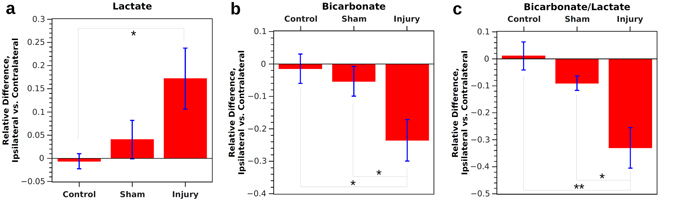
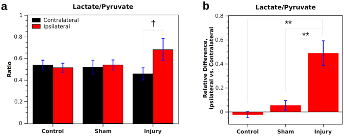
Traumatic brain injury (TBI) is known to cause perturbations in the energy metabolism of the brain, but current tests of metabolic activity are only indirect markers of energy use or are highly invasive. Here we show that hyperpolarized 13C magnetic resonance spectroscopic imaging (MRSI) can be used as a direct, non-invasive method for studying the effects of TBI on energy metabolism. Measurements were performed on rats with moderate TBI induced by controlled cortical impact on one cerebral hemisphere. Following injection of hyperpolarized [1-13C]pyruvate, the resulting 13C-bicarbonate signal was found to be 24 ± 6% lower in the injured hemisphere compared with the non-injured hemisphere, while the hyperpolarized bicarbonate-to-lactate ratio was 33 ± 8% lower in the injured hemisphere. In a control group, no significant difference in signal was found between sides of the brain. The results suggest an impairment in mitochondrial pyruvate metabolism, resulting in a decrease in aerobic respiration at the location of injury following TBI.


Conflict of interest statement

The authors declare that they have no competing interests.

Figures

Figure 1
Figure 1
(a) T2-weighted proton image showing diffuse injury to the cerebral cortex following CCI injury. The ipsilateral and contralateral regions of interest (ROIs) are shown by red and green outlines, respectively. (b) and (c) Fluoro-Jade B staining of the ipsilateral and contralateral hemispheres, respectively, 30 days after CCI injury. Fenestrated, dead tissue is outlined in orange and dead or dying cells are outlined in green. (d) Two typical hyperpolarized 13C MRSI spectra taken from ROIs on either side of the brain in an injured animal, showing an increase in lactate (lac) and decrease in bicarbonate (bic) intensity on the ipsilateral side. Pyruvate (pyr) and pyruvate hydrate (pyh) are also shown. (e) and (f) Maps of 13C signal for pyruvate (pyr), lactate (lac), bicarbonate (bic), and the bicarbonate-to-lactate ratio (bic/lac) in the rat brain of a control animal and an animal injured with a 2.0 mm deep controlled cortical impact (CCI), respectively. Significant differences in lac, bic, and bic/lac are evident in the brain surrounding the site of injury versus the rest of the brain. The control animal shows a more symmetric signal distribution.
Figure 2
Figure 2
Integrated signals from ipsilateral and contralateral ROIs in injured, sham, and control rats. Absolute signal intensity is arbitrary but signals for all metabolites are on the same scale. For pyruvate (a), the signal is similar for all cases. Lactate signal (b) is significantly lower on the contralateral side of sham and CCI animals. Bicarbonate signal (c) is significantly lower on the injured side of the brain following CCI, both compared with controls and the contralateral side. Bicarbonate-to-lactate ratio (d) is significantly lower on the ipsilateral than the contralateral side of the brain in both sham and CCI animals. *Indicates p < 0.05 and ** indicates p < 0.01 for independent samples. Indicates p < 0.05 for correlated samples.
Figure 3
Figure 3
The relative difference between ipsilateral and contralateral sides within control, sham, and injured animals. For lactate (a), control and sham animals have a similar signal on both sides of the brain, whereas injured animals have 17 ± 7% higher signal on the injured side. For bicarbonate (b), control and sham animals have a similar signal on both sides of the brain, whereas injured animals have 24 ± 6% lower signal on the injured side. For the bicarbonate-to-lactate ratio (c), control and sham animals have a similar ratio on each side of the brain, while injured animals have a 33 ± 8% lower ratio on the injured side. *Indicates p < 0.05 and **indicates p < 0.01 for independent samples.
Figure 4
Figure 4
Results for lactate-to-pyruvate ratio in injured, sham, and control rats. (a) Integrated values from ipsilateral and contralateral ROIs are similar for sham and control rats. For injured rats, the value is significantly higher on the ipsilateral vs. contralateral side (correlated t-test). (b) When considering the relative difference between ipsilateral and contralateral sides, control and sham animals have a similar value on both sides of the brain. Injured animals have a 49 ± 10% higher value on the injured side. **Indicates p < 0.01 for independent samples. Indicates p < 0.05 for correlated samples.

References

    1. Coronado, V. G., McGuire, L. C., Faul, M. F., Sugerman, D. E. & Pearson, W. S. In Brain Injury Medicine: Principles and Practice, Zasler, N. D., Katz, D. I. & Zafonte, R. D. editors, 84–100. Demos Medical, New York (2012).
    1. Roozenbeek B, Maas AIR, Menon DK. Changing patterns in the epidemiology of traumatic brain injury. Nat. Rev. Neurol. 2013;9:231–236. doi: 10.1038/nrneurol.2013.22. - DOI - PubMed
    1. Kolias AG, Guilfoyle MR, Helmy A, Allanson J, Hutchinson PJ. Traumatic brain injury in adults. Pract. Neurol. 2013;13:228–235. doi: 10.1136/practneurol-2012-000268. - DOI - PubMed
    1. McKenna MC, Scafidi S, Robertson CL. Metabolic Alterations in Developing Brain After Injury: Knowns and Unknowns. Neurochem. Res. 2015;40:2527–2543. doi: 10.1007/s11064-015-1600-7. - DOI - PMC - PubMed
    1. Jalloh I, et al. Lactate uptake by the injured human brain: evidence from an arteriovenous gradient and cerebral microdialysis study. J. Neurotrauma. 2013;30:2031–2037. doi: 10.1089/neu.2013.2947. - DOI - PMC - PubMed

Publication types