Review
. 2017 May 1:10:120.
doi: 10.3389/fnmol.2017.00120. eCollection 2017.

PINK1/Parkin-Dependent Mitochondrial Surveillance: From Pleiotropy to Parkinson's Disease

Affiliations
Review

PINK1/Parkin-Dependent Mitochondrial Surveillance: From Pleiotropy to Parkinson's Disease

Francois Mouton-Liger et al. Front Mol Neurosci. .

Abstract

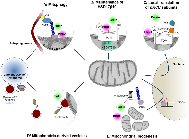
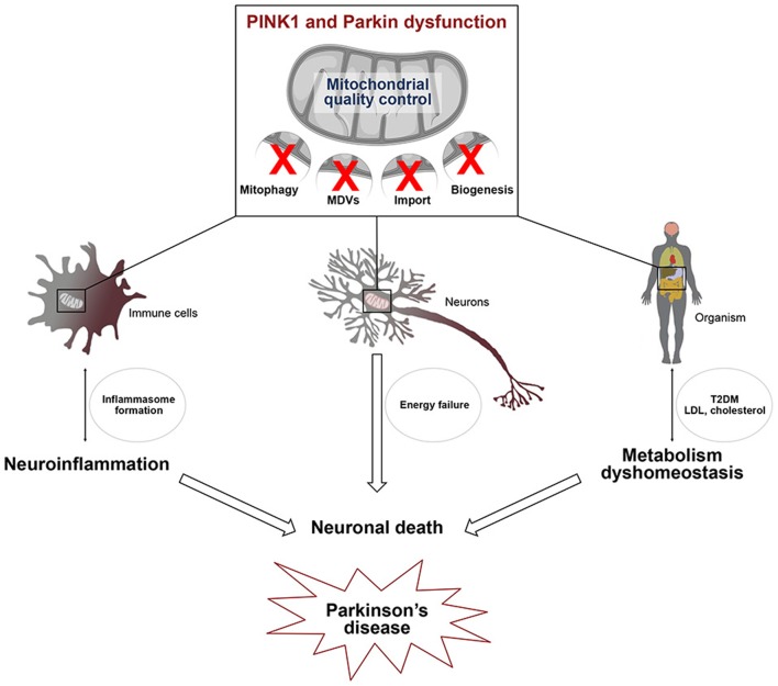
Parkinson's disease (PD) is one of the most frequent neurodegenerative disease caused by the preferential, progressive degeneration of the dopaminergic (DA) neurons of the substantia nigra (SN) pars compacta. PD is characterized by a multifaceted pathological process involving protein misfolding, mitochondrial dysfunction, neuroinflammation and metabolism deregulation. The molecular mechanisms governing the complex interplay between the different facets of this process are still unknown. PARK2/Parkin and PARK6/PINK1, two genes responsible for familial forms of PD, act as a ubiquitous core signaling pathway, coupling mitochondrial stress to mitochondrial surveillance, by regulating mitochondrial dynamics, the removal of damaged mitochondrial components by mitochondria-derived vesicles, mitophagy, and mitochondrial biogenesis. Over the last decade, PINK1/Parkin-dependent mitochondrial quality control emerged as a pleiotropic regulatory pathway. Loss of its function impinges on a number of physiological processes suspected to contribute to PD pathogenesis. Its role in the regulation of innate immunity and inflammatory processes stands out, providing compelling support to the contribution of non-cell-autonomous immune mechanisms in PD. In this review, we illustrate the central role of this multifunctional pathway at the crossroads between mitochondrial stress, neuroinflammation and metabolism. We discuss how its dysfunction may contribute to PD pathogenesis and pinpoint major unresolved questions in the field.

Keywords: Parkinson disease; Pink1/Parkin; UPRmt; mitochondrial quality control; mitochondrial stress; neuroinflammation; neuronal death.


Figures

Figure 1
Figure 1
Mitochondrial quality control mechanisms regulated by PINK1 and Parkin. PINK1 and Parkin have cooperative roles in several processes implicated in mitochondrial surveillance. (A) When mitochondria are damaged, Parkin is recruited to the mitochondrial surface and activated by PINK1, to exert its E3 ubiquitin protein ligase function. Ubiquitin chains on mitochondria are preferentially bound by sequestosome 1/p62-like autophagy receptors (SLRs), such as Optineurin and NDP52, which in turn mediate autophagosome formation on the damaged mitochondrion, leading to mitophagy. (B) Parkin is also involved in maintenance of mitochondrial levels of the multifunctional PD-linked mitochondrial enzyme HSD17B10, possibly by promoting its import through the TOM complex (C) PINK1 and Parkin can also control localized translation of several mRNAs for nuclear-encoded subunits of respiratory chain complexes (nRCC). Translationally repressed nRCC mRNAs are localized in a PINK1/Tom20-dependent manner to the mitochondrial outer membrane, where they are derepressed by Parkin through ubiquitin-dependent displacement of translation repressors, including Pumilio and Glorund/hnRNP-F. (D) PINK1 and Parkin also control the formation of so-called mitochondria-derived vesicles (MDVs), leading to the specific degradation of oxidized or damaged mitochondrial content by late endosomes/lysosomes. In this process, Syntaxin-17 is recruited to MDVs during budding, forming a ternary SNARE complex at the late endosome, together with SNAP29 and VAMP7, to mediate fusion. (E) Parkin also regulates the process of mitochondrial biogenesis by promoting the proteasomal degradation of the PGC-1α transcriptional repressor PARIS, in a PINK1-dependent manner.
Figure 2
Figure 2
Defects in mitochondrial quality control affect several pathways linked to Parkinson's disease. PINK1/Parkin-dependent mitochondrial quality control, including mitophagy, the MDV pathway and mitochondrial biogenesis, is essential for cell viability. Alterations of this process in several cell types may be linked to 1/ microglial activation leading to the transcriptional induction of genes encoding components of the NLRP3 inflammasome and IL-1β 2/ energy failure in neurons, due to the presence of damaged mitochondria and 3/ type 2 diabetes (T2DM) and low density lipoprotein (LDL) cholesterol accumulation. Activation of the innate immune system, metabolism dyshomeostasis and energetic defects contribute to neuronal death which is closely linked to Parkinson's disease.

References

    1. Anand R., Wai T., Baker M. J., Kladt N., Schauss A. C., Rugarli E., et al. . (2014). The i-AAA protease YME1L and OMA1 cleave OPA1 to balance mitochondrial fusion and fission. J. Cell Biol. 204, 919–929. 10.1083/jcb.201308006 - DOI - PMC - PubMed
    1. Arnould T., Michel S., Renard P. (2015). Mitochondria retrograde signaling and the UPR mt: where are we in mammals? Int. J. Mol. Sci. 16, 18224–18251. 10.3390/ijms160818224 - DOI - PMC - PubMed
    1. Ashrafi G., Schlehe J. S., LaVoie M. J., Schwarz T. L. (2014). Mitophagy of damaged mitochondria occurs locally in distal neuronal axons and requires PINK1 and Parkin. J. Cell Biol. 206, 655–670. 10.1083/jcb.201401070 - DOI - PMC - PubMed
    1. Ashrafi G., Wu Z., Farrell R. J., Ryan T. A. (2017). GLUT4 mobilization supports energetic demands of active synapses. Neuron 93, 606.e3–615.e3. 10.1016/j.neuron.2016.12.020 - DOI - PMC - PubMed
    1. Baker B. M., Nargund A. M., Sun T., Haynes C. M. (2012). Protective coupling of mitochondrial function and protein synthesis via the eIF2α kinase GCN-2. PLoS Genet. 8:e1002760. 10.1371/journal.pgen.1002760 - DOI - PMC - PubMed

LinkOut - more resources