. 2017 Apr 1;8(4):3192-3203.
doi: 10.1039/c6sc05720a. Epub 2017 Feb 8.

ANI-1: an extensible neural network potential with DFT accuracy at force field computational cost

Affiliations

ANI-1: an extensible neural network potential with DFT accuracy at force field computational cost

J S Smith et al. Chem Sci. .

Abstract

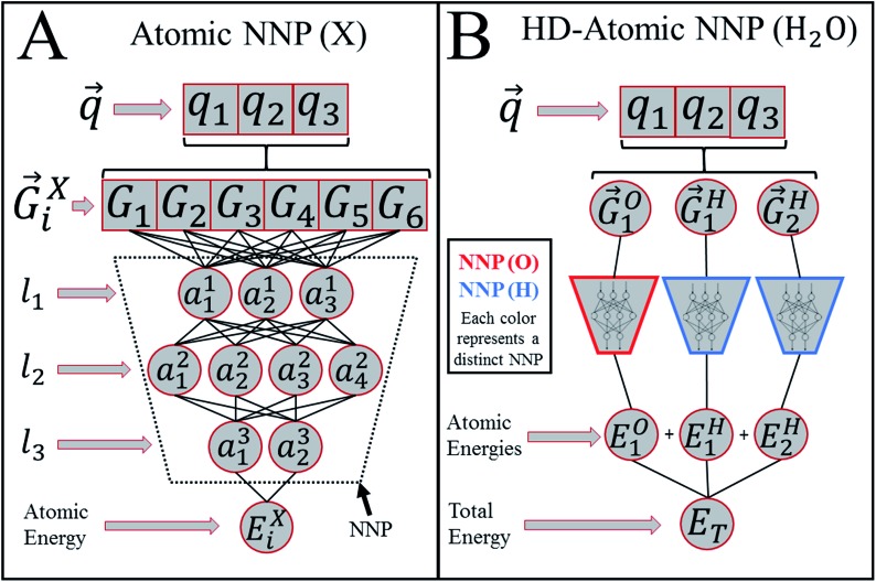
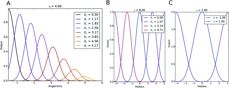
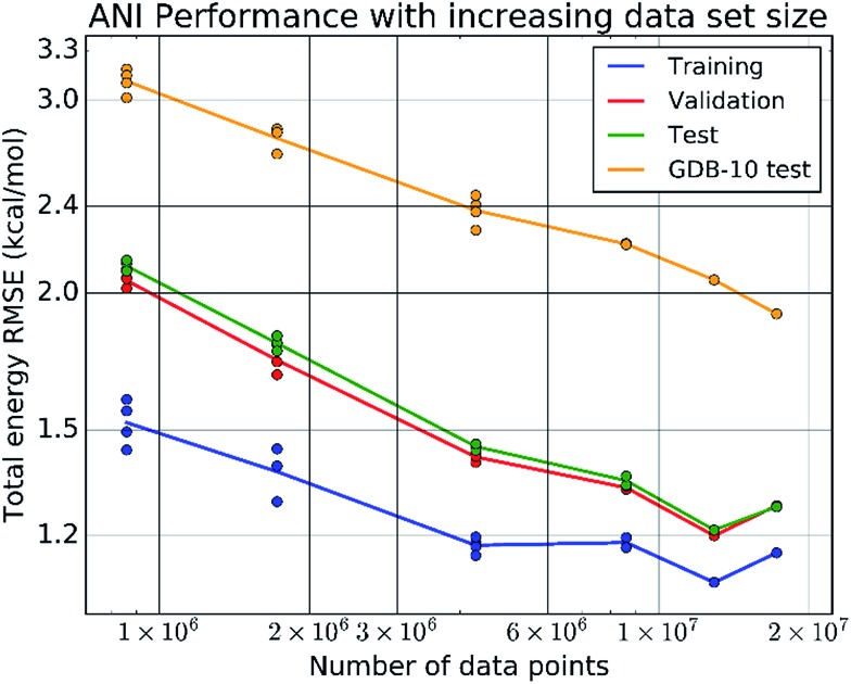
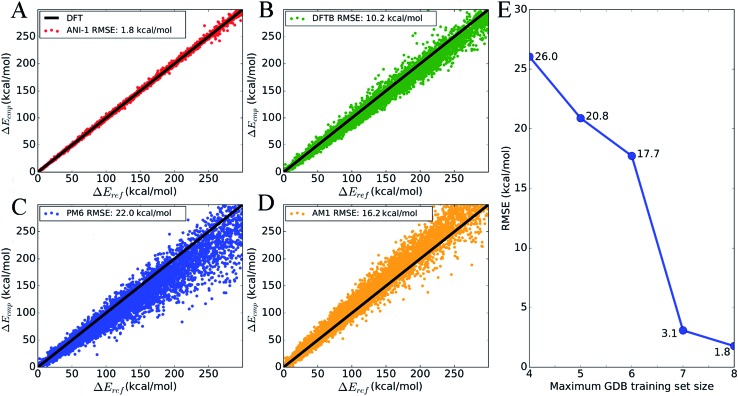
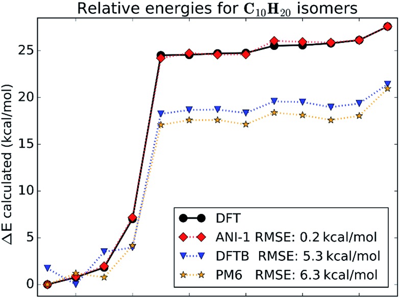
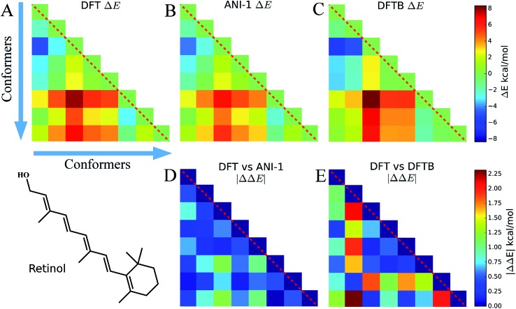
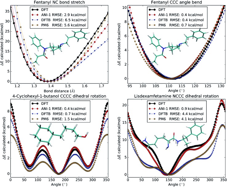
Deep learning is revolutionizing many areas of science and technology, especially image, text, and speech recognition. In this paper, we demonstrate how a deep neural network (NN) trained on quantum mechanical (QM) DFT calculations can learn an accurate and transferable potential for organic molecules. We introduce ANAKIN-ME (Accurate NeurAl networK engINe for Molecular Energies) or ANI for short. ANI is a new method designed with the intent of developing transferable neural network potentials that utilize a highly-modified version of the Behler and Parrinello symmetry functions to build single-atom atomic environment vectors (AEV) as a molecular representation. AEVs provide the ability to train neural networks to data that spans both configurational and conformational space, a feat not previously accomplished on this scale. We utilized ANI to build a potential called ANI-1, which was trained on a subset of the GDB databases with up to 8 heavy atoms in order to predict total energies for organic molecules containing four atom types: H, C, N, and O. To obtain an accelerated but physically relevant sampling of molecular potential surfaces, we also proposed a Normal Mode Sampling (NMS) method for generating molecular conformations. Through a series of case studies, we show that ANI-1 is chemically accurate compared to reference DFT calculations on much larger molecular systems (up to 54 atoms) than those included in the training data set.


Figures

Fig. 1
Fig. 1. Behler and Parrinello's HDNN or HD-atomic NNP model. (A) A scheme showing the algorithmic structure of an atomic number specific neural network potential (NNP). The input molecular coordinates, q, are used to generate the atomic environment vector, G i X, for atom i with atomic number X. G i X is then fed into a neural network potential (NNP) trained specifically to predict atomic contributions, E i X, to the total energy, E T. Each l k represents a hidden layer of the neural network and is composed of nodes denoted a j k where j indexes the node. (B) The high-dimensional atomic NNP (HD-atomic NNP) model for a water molecule. G i X is computed for each atom in the molecule then input into their respective NNP (X) to produce each atom's E i X, which are summed to give E T.
Fig. 2
Fig. 2. Examples of the symmetry functions with different parameter sets. (A) Radial symmetry functions, (B) modified angular symmetry functions and (C) the original Behler and Parrinello angular symmetry functions. These figures all depict the use of multiple shifting parameters for each function, while keeping the other parameters constant.
Fig. 3
Fig. 3. Log–log plots of the training, validation, testing, and a random GDB-10 (molecules with 10 heavy atoms from the GDB-11 database) extensibility testing set of total energy errors vs. increasing number of data points in the training set. The sets of points converge to the final ANI-1 potential presented in this paper, trained on the full ANI-1 data set.
Fig. 4
Fig. 4. Relative energy comparisons from random conformations of a random sampling of 134 molecules from GDB-11 all with 10 heavy atoms. There is an average of 62 conformations, and therefore energies, per molecule. Each set of energies for each molecule is shifted such that the lowest energy is at 0. None of the molecules from this set are included in any of the ANI training sets. (A–D) Correlation plots between DFT energies, E ref, and computed energies, E cmp, for ANI-1 and popular semi-empirical QM methods. Each individual molecule's set of energies is shifted such that the lowest energy is at zero. (E) RMS error (kcal mol–1) of various ANI potentials, compared to DFT, trained to an increasing data set size. The x-axis represents the maximum size of GDB molecules included in the training set. For example, 4 represents an ANI potential trained to a data set built from the subset of GDB-11 containing all molecules up to 4 heavy atoms.
Fig. 5
Fig. 5. The total energies, shifted such that the lowest is zero, calculated for various C10H20 isomers, are compared between DFT with the ωB97X functional and 6-31G(d) basis set, the ANI-1 potential, AM1 semi-empirical, and PM6 semi-empirical methods.
Fig. 6
Fig. 6. (A–C) These three triangle plots, which are on the same scale shown to the right, show energy differences, ΔE, between random energy minimized conformers of the molecule retinol. The structural differences between these conformers include many dihedral rotations. (A) shows the conformers ΔE calculated with DFT, (B) ANI-1, and (C) DFTB. (D) shows the absolute value of the difference between (A) and (B), |ΔΔE|, while (E) shows the same between (A) and (C). ΔE and |ΔΔE| have their own scale shown to the right of the plots. All plots of a specific type use the same color scaling for easy comparison.
Fig. 7
Fig. 7. Each subplot shows a one-dimensional potential surface scan generated from DFT, the ANI-1 potential, and two popular semi-empirical methods, DFTB and PM6. The atoms used to produce the scan coordinate are labeled in the images of the molecules in every sub-plot. Each figure also lists the RMSE, in the legend, for each method compared to the DFT potential surface.

References

    1. Kitaura K., Ikeo E., Asada T., Nakano T., Uebayasi M. Chem. Phys. Lett. 1999;313:701–706.
    1. Fedorov D. G., Nagata T., Kitaura K. Phys. Chem. Chem. Phys. 2012;14:7562. - PubMed
    1. Ochsenfeld C., Kussmann J. and Lambrecht D. S., in Reviews in Computational Chemistry, John Wiley & Sons, Inc., 2007, pp. 1–82.
    1. Elstner M. Theor. Chem. Acc. 2006;116:316–325.
    1. Stewart J. J. P. J. Mol. Model. 2009;15:765–805. - PubMed