Review
. 2017 Apr 27;2(3):73-82.
doi: 10.1302/2058-5241.2.160069. eCollection 2017 Mar.

Spinal osteotomies: indications, limits and pitfalls

Affiliations
Review

Spinal osteotomies: indications, limits and pitfalls

Kamil Cagri Kose et al. EFORT Open Rev. .

Abstract

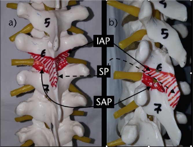
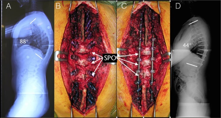
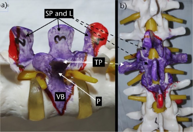
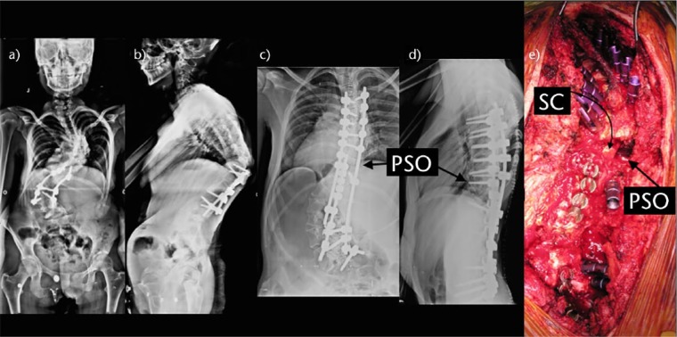
The aims of spinal deformity surgery are to achieve balance, relieve pain and prevent recurrence or worsening of the deformity.The main types of osteotomies are the Smith-Petersen osteotomy (SPO), pedicle subtraction osteotomy (PSO), bone-disc-bone osteotomy (BDBO) and vertebral column resection (VCR), in order of increasing complexity.SPO is a posterior column osteotomy in which the posterior ligaments and the facet joints are removed and correction is performed through the disc space. A mobile anterior disc is essential. SPO is best in patients with +6-8 cm C7 plumbline. The amount of correction is 9.3° to 10.7°/level (1°/mm bone).PSO is a technique where the posterior elements and pedicles are removed. Then a triangular wedge through the pedicles is removed and the posterior spine is shortened using the anterior cortex as a hinge. The ideal candidates are patients with a severe sagittal imbalance. A single level osteotomy can produce 30° 40° of correction. A single level osteotomy may restore global sagittal balance by an average of 9 cm with an upper limit of 19 cm.BDBO is an osteotomy done above and below a disc level. A BDBO provides correction rates in the range of 35° to 60°. The main indications are deformities with the disc space as the apex and severe sagittal plane deformities.VCR is indicated for rigid multi-planar deformities, sharp angulated deformities, hemivertebra resections, resectable spinal tumours, post-traumatic deformities and spondyloptosis. The main indication for a VCR is fixed coronal plane deformity.The type of osteotomy must be chosen mainly according to the aetiology, type and apex of the deformity. One may start with SPOs and may gradually advance to complex osteotomies. Cite this article: EFORT Open Rev 2017;2:73-82. DOI: 10.1302/2058-5241.2.160069.


Conflict of interest statement

ICMJE Conflict of Interest Statement: None

Figures

Fig. 1
Fig. 1
Smith-Petersen osteotomy. a) Spine from posterior view. The spinous process and facets of the upper vertebra are removed in addition to the superior facets of the inferior vertebra. The areas to be removed are painted in red. b) Spine from the lateral view. The areas to be resected are painted in red. the correction is based on the movement of the disc space. IAP, inferior articular process of the superior vertebra; SP, spinous process; SAP, superior articular process of the inferior vertebra.
Fig. 2
Fig. 2
Smith-Petersen osteotomies in a Scheuermann’s kyphosis patient. a) A 19-year-old male patient with Scheuermann’s kyphosis. Lateral radiograph. Thoracic kyphosis is 88°. b) Intra-operative view of T6-7, T7-8, T8-9, T9-10 Smith Petersen osteotomies. c) Intra-operative view after correction and stabilisation procedure. Note that the osteotomy sites were closed under compression. d) Lateral post-operative radiograph. The kyphosis angle is 44° after correction. SPO, Smith-Petersen osteotomy.
Fig. 3
Fig. 3
Pedicle subtraction osteotomy. a) An L3 pedicle subtraction osteotomy plan from the lateral view. Note that a wedge reaching to, but not crossing, the anterior cortex is to be removed. The parts of the laminae painted in red may be preserved if they do not interfere with closure of the osteotomy gap. b) While planning an osteotomy, laminectomy of the upper and lower vertebrae should be done to prevent buckling of the spinal cord after correction of the deformity. Again, the area painted in red may not be removed if enough decompression is done and these structures do not prevent closure of the osteotomy. SP, spinous process; L, lamina; TP, transverse process; VB, vertebral body; P, pedicle.
Fig. 4
Fig. 4
Pedicle subtraction osteotomy in revision scoliosis surgery. a, b) Anteroposterior (AP) and lateral views of a 21-year-old female patient who had undergone a previously unsuccessful fusion operation. She presented with both a severe scoliosis and also decompensated kyphosis. The dotted white line represents the planned osteotomy. c, d) AP and lateral views after surgery. There was a significant correction of both coronal and sagittal balance. The osteotomy sites are shown with black arrows. e) Intra-operative view of an asymmetric T10 pedicle subtraction osteotomy and instrumentation. Spinal cord and osteotomy site are shown with black arrows. SC, spinal cord; PSO, pedicle subtraction osteotomy.
Fig. 5
Fig. 5
Patient positioning in a severe kyphosis patient. In severe kyphosis, instead of using a four-poster frame, two separate posts (P in the picture) can be used. In this case, one post was placed under the sternum and the other was placed under the pelvis. Please note that the operating table was also bent to comply with the global kyphosis of the patient. P.
Fig. 6
Fig. 6
Bone-disc-bone osteotomy. a) Spine from the lateral view. Upper end-plate of T10 and lower end-plate of T9 were included in the area to be resected. Resection should also include the disc space. Closure of the osteotomy allows bone-to-bone contact and direct bone healing. b) Spine from the posterior view. Lower facets and spinous process of the upper vertebra and upper facets of the lower vertebra should be removed to gain access to the area to be resected. SP, spinous process; L, lamina; IF, inferior facet; SF, superior facet; PIC, postero-inferior corner; PSC, posterosuperior corner; D, disc.
Fig. 7
Fig. 7
Vertebral column resection. a) Lateral view. Lower facets of the upper and upper facets of the lower vertebrae should be removed to allow removal of the laminae and pedicles of the vertebra to be resected. Discs should also be resected and this should be followed by end-plate preparation for a successful fusion. b) AP view of the anatomical structures to be resected during a PVCR operation. IAP, inferior articular process; D, disc; SP, spinous process; SAP, superior articular process; VB, vertebral body; L, lamina.
Fig. 8
Fig. 8
a) A 33-year-old woman with congenital kyphosis. b) Intra-operative view of the kyphotic region. c) Intra-operative view of three levels. Vertebral column resection including T11, T12 and L1. d) Post-operative lateral view. AK, apex of kyphosis; L, lamina; SP, spinous process; NR, nerve root; SC, spinal cord.

References

    1. Smith-Petersen MN, Larson EB, AuFranc OE. Osteotomy of the spine for correction of flexion deformity in rheumatoid arthritis. J Bone Joint Surg [Am] 1945;27-A:1-11. - PubMed
    1. Ponte A, Vero B, Siccardi GL. Surgical treatment of Scheuermann’s kyphosis. In: Winter RB, ed. Progress in spinal pathology: kyphosis. Bologna: Aulo Gaggi, 1984:75-80.
    1. Bridwell KH. Decision making regarding Smith-Petersen vs. pedicle subtraction osteotomy vs. vertebral column resection for spinal deformity. Spine (Phila Pa 1976) 2006;31:S171-S178. - PubMed
    1. Dorward IG, Lenke LG. Osteotomies in the posterior-only treatment of complex adult spinal deformity: a comparative review. Neurosurg Focus 2010;28:E4. - PubMed
    1. Lafage V, Schwab F, Patel A, Hawkinson N, Farcy JP. Pelvic tilt and truncal inclination: two key radiographic parameters in the setting of adults with spinal deformity. Spine (Phila Pa 1976) 2009;34:E599-E606. - PubMed

LinkOut - more resources