. 2017 Feb 17;6(4):e1293210.
doi: 10.1080/2162402X.2017.1293210. eCollection 2017.

IL15 induces a potent antitumor activity in NK cells isolated from malignant pleural effusions and overcomes the inhibitory effect of pleural fluid

Affiliations

IL15 induces a potent antitumor activity in NK cells isolated from malignant pleural effusions and overcomes the inhibitory effect of pleural fluid

D Croxatto et al. Oncoimmunology. .

Abstract

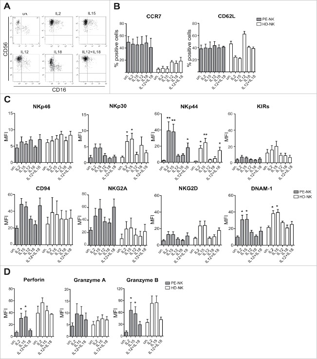
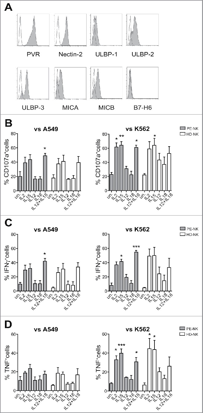
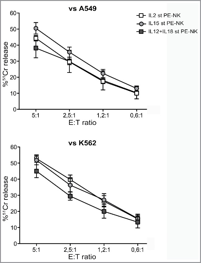
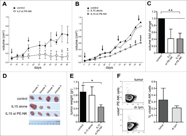
Natural Killer (NK) cells are capable of recognizing and killing cancer cells and play an important role in tumor immunosurveillance. However, tumor-infiltrating NK cells are frequently impaired in their functional capability. A remarkable exception is represented by NK cells isolated from malignant pleural effusions (PE) that are not anergic and, upon IL2-induced activation, efficiently kill tumor cells. Although IL2 is used in various clinical trials, severe side effects may occur in treated patients. In this study, we investigated whether also other clinical-grade cytokines could induce strong cytotoxicity in NK cells isolated from pleural fluid of patients with primary or metastatic tumors of different origins. We show that PE-NK cells, cultured for short-time intervals with IL15, maintain the CD56bright phenotype, a high expression of the main activating receptors, produce cytokines and kill tumor cells in vitro similarly to those treated with IL2. Moreover, IL15-activated PE-NK cells could greatly reduce the growth of established tumors in mice. This in vivo antitumor effect correlated with the ability of IL15-activated PE-NK cells to traffic from periphery to the tumor site. Finally, we show that IL15 can counteract the inhibitory effect of the tumor pleural microenvironment. Our study suggests that IL15-activated NK cells isolated from pleural fluid (otherwise discarded after thoracentesis) may represent a suitable source of effector cells to be used in adoptive immunotherapy of cancer.

Keywords: Adoptive immunotherapy; IL15; NK cells; innate lymphoid cells (ILC); pleural fluid; pleural tumors.


Figures

Figure 1.
Figure 1.
Cytokine-stimulation induces phenotypic changes in PE-NK cells. (A–D) Freshly isolated PE- (gray bars) and peripheral blood HD-NK (white bars) cells were stimulated with IL2, IL15, IL12, and IL18 alone or in combination, for 72 h (short-term) and then analyzed. (A) CD56 and CD16 surface expression on unstimulated (un.) and on cytokine-stimulated PE-NK cells. One representative experiment out of 12 performed. (B) Percentages ± SEM of CCR7 and CD62L expression (n = 4). (C) Mean fluorescence intensity (MFI) ± SEM of the major activating and inhibitory NK receptors (n = 8). (D) MFI ± SEM of perforin, granzymes A, and B (n = 4). (B–D) Statistical analysis was performed using one-way ANOVA Kruskal–Wallis test, (*) indicates the comparison between cytokine stimulated and unstimulated NK cells within the same group (PE-NK or HD-NK). Where not indicated, data were not statistically significant.
Figure 2.
Figure 2.
Cytokine stimulation upregulates PE-NK cell function. (A) Surface expression of cell ligands recognized by activating NK receptors on A549 tumor cell line. One representative experiment out of four performed. (B–D) NK cells isolated from PE (gray bars) or from peripheral blood of HD (white bars) were stimulated for 72 h (short-term) with IL2, IL15, IL12, and IL18, alone or in combination. (B) Percentages ± SEM of CD107+ NK cells upon exposure to A549 (n = 5) or K562 target cells (n = 6). (C, D) Percentages ± SEM of IFNγ+ or TNF+ NK cells upon exposure to A549 (n = 5) or K562 target cells (n = 6). (B–D) Statistical analysis was performed using one-way ANOVA Kruskal–Wallis test, (*) indicates the comparison between cytokine-stimulated and unstimulated cells within the same group (PE-NK or HD-NK). When not indicated, data were not statistically significant.
Figure 3.
Figure 3.
Short-term IL15-activated PE-NK cells display efficient cytolytic activity. Cytolytic activity of IL2, or IL15 or IL12+IL18 short-term (st) PE-NK cells was evaluated. Mean of 51Cr release ± SEM using A549 (n = 6–8) or K562 (n = 5–6) as target cells. The Effector:Target (E:T) ratios are indicated.
Figure 4.
Figure 4.
IL15-activated PE-NK cells controlled tumor growth in vivo. (A–F) A549 were injected subcutaneously (s.c.) into athymic nude mice (day 0). At different time intervals mice were treated with PBS (control), or IL15 alone (IL15), or with PE-NK cells pre-activated by IL2- or IL15-short-term (st) culture (72h). (A) Mice were treated every 11 d (see arrows) with IL2 st PE-NK cells (white symbols) derived from same patient or with PBS (black symbols), starting when tumors were palpable (day 10). Data indicate the mean tumor volume ± SEM (n = 4 per group).(B) Mice were treated every 11 d (see arrows) with IL15 st PE-NK cells (gray symbols, n = 4) derived from same patient, or with IL15 alone (white symbols, n = 3), or with PBS (black symbols, n = 4), starting when tumors were palpable (day 11). Data indicate the mean tumor volume. (A, B) Statistical analysis was performed using two-way ANOVA test, (*) indicates the comparison between mice infused with IL2- or IL15-short-term PE-NK cells and the corresponding PBS-treated mice group .Where not indicated, data are not statistically significant. (C) Fold change ± SEM of tumor volume (at day 39) of IL2- or IL15-short-term PE-NK cells treated group calculated on the basis of the tumor volume detected in control group (PBS), arbitrarily normalized to 1. Statistical analysis was performed using Mann–Whitney test, comparing treated mice with control group. Where not indicated, data are not statistically significant.(D) Representative image of tumors isolated from mice at day 45, each treatment group is indicated. (E) Mean tumor weight ± SEM was measured at day 45 in groups of mice treated as indicated. Statistical analysis was performed using Mann–Whitney test, comparing mice treated with IL15-activated PE-NK cells or IL15 alone with control group. When not indicated, data were not statistically significant. (F) Recruitment of IL15 st PE-NK cells in tumors and tumor draining lymph nodes (dr.lym.) in the corresponding treated mice. One representative experiment and percentages ± SEM of violet+ NK cells recovered (n = 4).
Figure 5.
Figure 5.
Soluble factors detected in malignant pleural effusions and in autologous tumor cell line supernatants. Malignant pleural effusion and their autologous tumor cell line culture supernatants were analyzed by ELISA and Luminex multiplex assays. Mean ± SEM of cytokine concentration (pg/mL) (n = 5).
Figure 6.
Figure 6.
IL15 counteracts the inhibitory effect of malignant pleural fluid. (A, B) IL15 short-term (st) PE-NK cells were cultured under different culture conditions and their cytolytic activity against A549 cells was analyzed (E:T ratio, 5:1). Pre-activated IL15 st PE-NK cells (white bar) were washed and subsequently: (A) cultured with autologous PE for different time intervals (2 h, 24 h, and 72 h) (gray bars) (n = 5); (B) cultured for 72 h with the following: autologous PE (dark gray bar), RPMI (light gray bar), autologous PE+IL15 (dark gray stripped bar), and RPMI+IL15 (light gray stripped bar) (n = 3–7). (C) Pre-activated IL15 st PE-NK cells cultured subsequently for 72 h with autologous PE (dark gray bar) were washed and cultured for additionally 72 h with IL15 and autologous PE or RPMI (dark and light gray stripped bars, respectively) (n = 3). (A–C) Percentages ± SEM of 51Cr release. Statistical analysis was performed using Mann–Whitney test. Where not indicated data were not statistically significant.

References

    1. Spits H, Artis D, Colonna M, Diefenbach A, Di Santo JP, Eberl G, Koyasu S, Locksley RM, McKenzie AN, Mebius RE et al.. Innate lymphoid cells–a proposal for uniform nomenclature. Nat Rev Immunol 2013; 13:145-9; PMID:23348417; http://dx.doi.org/10.1038/nri3365 - DOI - PubMed
    1. Montaldo E, Vacca P, Moretta L, Mingari MC. Development of human natural killer cells and other innate lymphoid cells. Semin Immunol 2014; 26:107-13; PMID:24559836; http://dx.doi.org/10.1016/j.smim.2014.01.006 - DOI - PubMed
    1. Eberl G, Di Santo JP, Vivier E. The brave new world of innate lymphoid cells. Nat Immunol 2015; 16:1-5; PMID:25521670; http://dx.doi.org/10.1038/ni.3059 - DOI - PubMed
    1. Vivier E, Raulet DH, Moretta A, Caligiuri MA, Zitvogel L, Lanier LL, Yokoyama WM, Ugolini S. Innate or adaptive immunity? The example of natural killer cells. Science 2011; 331:44-9; PMID:21212348; http://dx.doi.org/10.1126/science.1198687 - DOI - PMC - PubMed
    1. Moretta A, Bottino C, Vitale M, Pende D, Biassoni R, Mingari MC, Moretta L. Receptors for HLA class-I molecules in human natural killer cells. Annu Rev Immunol 1996; 14:619-48; PMID:8717527; http://dx.doi.org/10.1146/annurev.immunol.14.1.619 - DOI - PubMed

Publication types