. 2017 Mar 3;6(4):e1295903.
doi: 10.1080/2162402X.2017.1295903. eCollection 2017.

Preclinical efficacy of immune-checkpoint monotherapy does not recapitulate corresponding biomarkers-based clinical predictions in glioblastoma

Affiliations

Preclinical efficacy of immune-checkpoint monotherapy does not recapitulate corresponding biomarkers-based clinical predictions in glioblastoma

Abhishek D Garg et al. Oncoimmunology. .

Abstract

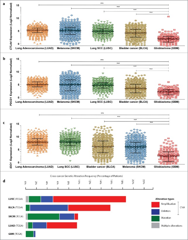
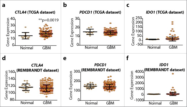
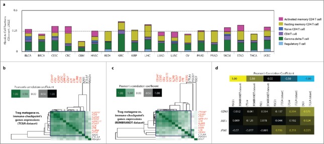
Glioblastoma (GBM) is resistant to most multimodal therapies. Clinical success of immune-checkpoint inhibitors (ICIs) has spurred interest in applying ICIs targeting CTLA4, PD1 or IDO1 against GBM. This amplifies the need to ascertain GBM's intrinsic susceptibility (or resistance) toward these ICIs, through clinical biomarkers that may also "guide and prioritize" preclinical testing. Here, we interrogated the TCGA and/or REMBRANDT human patient-cohorts to predict GBM's predisposition toward ICIs. We exploited various broad clinical biomarkers, including mutational or predicted-neoantigen burden, pre-existing or basal levels of tumor-infiltrating T lymphocytes (TILs), differential expression of immune-checkpoints within the tumor and their correlation with particular TILs/Treg-associated functional signature and prognostic impact of differential immune-checkpoint expression. Based on these analyses, we found that predictive biomarkers of ICI responsiveness exhibited inconsistent patterns in GBM patients, i.e., they either predicted ICI resistance (as compared with typical ICI-responsive cancer-types like melanoma, lung cancer or bladder cancer) or susceptibility to therapeutic targeting of CTLA4 or IDO1. On the other hand, our comprehensive literature meta-analysis and preclinical testing of ICIs using an orthotopic GL261-glioma mice model, indicated significant antitumor properties of anti-PD1 antibody, whereas blockade of IDO1 or CTLA4 either failed or provided very marginal advantage. These trends raise the need to better assess the applicability of ICIs and associated biomarkers for GBM.

Keywords: CTLA4; Cancer immunotherapy; IDO1; PD1; T cells; Treg cells; copy-number alterations (CNA); high-grade glioma; immune-checkpoint blockers (ICBs); mutational burden; neoantigen; patient prognosis.


Figures

Figure 1.
Figure 1.
Glioblastoma (GBM) has one of the lowest overall mutational and predicted neoantigen burdens. (A) Presence or absence of carcinogen-induced mutational signatures (derived from COSMIC-database: http://cancer.sanger.ac.uk/cosmic/signatures) was used as a means to “classify” median (somatic) mutational burdens of 18 TCGA cancer-data sets (mean ± s.d., Mann–Whitney test; p-value as indicated); (B) Correlation between median mutational burdens of different TCGA cancer-data sets and median mutational counts of corresponding cell lines belonging to these cancer types from the CCLE-data sets. (C) Correlation of median mutational burdens and median-predicted neoantigen burden from the respective TCGA data sets derived from The Cancer Immunome Atlas at http://tcia.at. Abbreviations: BLCA, bladder urothelial carcinoma; BRCA, breast cancer; CESC, cervical squamous cell carcinoma; CRC, colorectal carcinoma; GBM, glioblastoma; HNSC, head and neck squamous cell carcinoma; KICH, kidney chromophobe cancer; KIRC, kidney renal clear cell carcinoma; KIRP, kidney renal papillary cell carcinoma; LIHC, liver hepatocellular carcinoma; LUAD, lung adenocarcinoma; LUSC, lung squamous cell carcinoma; OV, ovarian cancer; PAAD, pancreatic adenocarcinoma; PRAD, prostate adenocarcinoma; SKCM, skin cutaneous melanoma; STAD, stomach adenocarcinoma; THCA, thyroid cancer; UCEC, uterine corpus endometrial carcinoma; Of note, mutational burden refers to nonsynonymous somatic single nucleotide variations.
Figure 2.
Figure 2.
Melanoma, lung cancer and bladder cancer exhibit significantly higher expression of CTLA4, PDCD1 or IDO1 than glioblastoma in human patients. Analysis of differential expression of CTLA4 (A), PDCD1 (B) or IDO1 (C) between lung adenocarcinoma (LUAD; n = 517), melanoma (SKCM; n = 472), lung squamous cell carcinoma (SCC) (LUSC; n = 501), bladder cancer (BLCA; n = 408) and glioblastoma (GBM; n = 166) (from TCGA cohorts) (data are log2 normalized and presented as median with inter-quartile range; One-way ANOVA; significance set at p < 0.05; ***p < 0.001). Of note RNASeq, and not microarray, expression data was used for this analysis since comprehensive microarray data are not available for melanoma and bladder cancer within TCGA data set. (D) A combined cross-cancer genetic alteration frequency analysis (expressed as % of patients) for CTLA4, PDCD1 and IDO1 genes was performed using the TCGA cohorts of LUAD, LUSC, SKCM, BLCA and GBM. The graph indicates presence of either genetic mutations or specific copy-number alterations (CNA; like genetic deletion or amplification) or presence of multiple such alterations simultaneously (as indicated by the color-code in the legend within the figure).
Figure 3.
Figure 3.
Glioblastoma (GBM) tumor tissue does not show strong upregulation of CTLA4, PDCD1 and IDO1. Analysis of differential expression of CTLA4 (A, D), PDCD1 (B, E) and IDO1 (C, F) between normal brain tissue sample (for REMBRANDT, n = 21; for TCGA, n = 11) and GBM tissue sample (for REMBRANDT, n = 214; for TCGA, n = 202) (mean ± s.d., Mann–Whitney test; p-value as indicated). Of note, TCGA and REMBRANDT data sets were analyzed by different expression-analysis platforms and standardized by different post-processing and normalization analyses and hence their overall gene-expression counts have different (but proportional) scales.
Figure 4.
Figure 4.
Glioblastoma (GBM) exhibits sparse basal T cell-infiltrates and correlation of CTLA4 and IDO1 with T-cell-associated polarization biomarkers. (A) The overall (absolute) presence of different T-cells-associated genetic signatures or metagenes (indicated as color code) was estimated across 19 TCGA cancer-data sets using The Cancer Immunome Atlas. (B, C, D) Correlation of CTLA4, PDCD1, IDO1 expression with, GBM-specific Treg-metagene across TCGA (B) and REMBRADT data sets (C) (blue-box indicates co-clustering of immune checkpoint with Treg-metagene); or with, GZMA, PRF1 and IFNG across the same data sets (D). BLCA, bladder urothelial carcinoma; BRCA, breast cancer; CESC, cervical squamous cell carcinoma; CRC, colorectal carcinoma; GBM, glioblastoma; HNSC, head and neck squamous cell carcinoma; KICH, kidney chromophobe cancer; KIRC, kidney renal clear cell carcinoma; KIRP, kidney renal papillary cell carcinoma; LIHC, liver hepatocellular carcinoma; LUAD, lung adenocarcinoma; LUSC, lung squamous cell carcinoma; OV, ovarian cancer; PAAD, pancreatic adenocarcinoma; PRAD, prostate adenocarcinoma; SKCM, skin cutaneous melanoma; STAD, stomach adenocarcinoma; THCA, thyroid cancer; UCEC, uterine corpus endometrial carcinoma.
Figure 5.
Figure 5.
CTLA4, PDCD1 and IDO1 differential expression does not exhibit significant prognostic impact in GBM patients. TCGA GBM-cohort (n = 540, A–C) and REMBRANDT GBM-cohort (n = 178, D–F) stratified (median) into “high-expression” (red; TCGA, n = 271; REMBRANDT, n = 89) or “low-expression” (black; TCGA, n = 269; REMBRANDT, n = 89) and represented as Kaplan–Meier plots (log-rank (Mantel-Cox) test; hazard ratios (HR)+95% confidence interval or CI).
Figure 6.
Figure 6.
Anti-PD1 mono-immunotherapy exhibits the highest therapeutic efficacy against preclinical glioma. (A, B) Mice inoculated with GL261 cells intra-axially, were treated with IDO1-inhibitors (A) or anti-CTLA4, anti-PD1 antibodies (B). Kaplan–Meier survival curves for glioma-bearing mice treated with IDO1 inhibitors (CNTR, n = 13; +1-D-MT, n = 15; +1-DL-MT, n = 5) (C), Ido1+/+ (n = 14) vs. Ido1−/− (n = 17) mice (D) or treated with anti-CTLA4 (IgG Ab, n = 9; CTLA4 Ab, n = 10) (E) or anti-PD1 (IgG Ab, n = 8; PD1 Ab, n = 12) (F) antibodies (Log-rank-(Mantel-Cox)-test). (G–L) C57BL/6 mice were inoculated with live GL261 cells (Day 0), intra-axially and either injected with respective IgG antibodies (Ab), i.e., control mice or treated with anti-CTLA4 Ab (G–I) or anti-PD1 Ab (J–L). Thereafter the mice were killed at day 18–22 post-GL261 inoculation and the brains were isolated. Initially, total mononuclear immune cells were counted. Thereafter these were processed for FACS-based immunophenotyping for (G) CTLA4+CD4+T cells, (H) CTLA4+CD8+T cells, (I) CTLA4+Treg cells, (J) PD1+CD4+T cells, (K) PD1+CD8+T cells and (L) PD1+Treg cells. The histograms are representative of n = 3–4 mice/group (the percentage of CTLA4 or PD1 negative and positive T cells are indicated through the agency of histogram).

Comment in

References

    1. Johanns TM, Ward JP, Miller CA, Wilson C, Kobayashi DK, Bender D, Fu Y, Alexandrov A, Mardis ER, Artyomov MN et al.. Endogenous neoantigen-specific CD8 T cells identified in two glioblastoma models using a cancer immunogenomics approach. Cancer Immunol Res 2016; 4:1007-15; PMID:27799140; http://dx.doi.org/10.1158/2326-6066.CIR-16-0156 - DOI - PMC - PubMed
    1. Preusser M, Lim M, Hafler DA, Reardon DA, Sampson JH. Prospects of immune checkpoint modulators in the treatment of glioblastoma. Nat Rev Neurol 2015; 11:504-14; PMID:26260659; http://dx.doi.org/10.1038/nrneurol.2015.139 - DOI - PMC - PubMed
    1. Polyzoidis S, Tuazon J, Brazil L, Beaney R, Al-Sarraj ST, Doey L, Logan J, Hurwitz V, Jarosz J, Bhangoo R et al.. Active dendritic cell immunotherapy for glioblastoma: Current status and challenges. Br J Neurosurg 2015; 29:197-205; PMID:25541743; http://dx.doi.org/10.3109/02688697.2014.994473 - DOI - PubMed
    1. Anguille S, Smits EL, Lion E, van Tendeloo VF, Berneman ZN. Clinical use of dendritic cells for cancer therapy. Lancet Oncol 2014; 15:e257-67; PMID:24872109; http://dx.doi.org/10.1016/S1470-2045(13)70585-0 - DOI - PubMed
    1. Garg AD, Vandenberk L, Koks C, Verschuere T, Boon L, Van Gool SW, Agostinis P. Dendritic cell vaccines based on immunogenic cell death elicit danger signals and T cell-driven rejection of high-grade glioma. Sci Transl Med 2016; 8:328ra27; http://dx.doi.org/10.1126/scitranslmed.aae0105 - DOI - PubMed

Publication types

LinkOut - more resources