

With Friends Like These: The Complex Role of Neutrophils in the Progression of Severe Pneumonia
- PMID: 28507954
- PMCID: PMC5410563
- DOI: 10.3389/fcimb.2017.00160
With Friends Like These: The Complex Role of Neutrophils in the Progression of Severe Pneumonia
Abstract
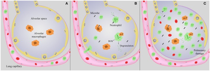
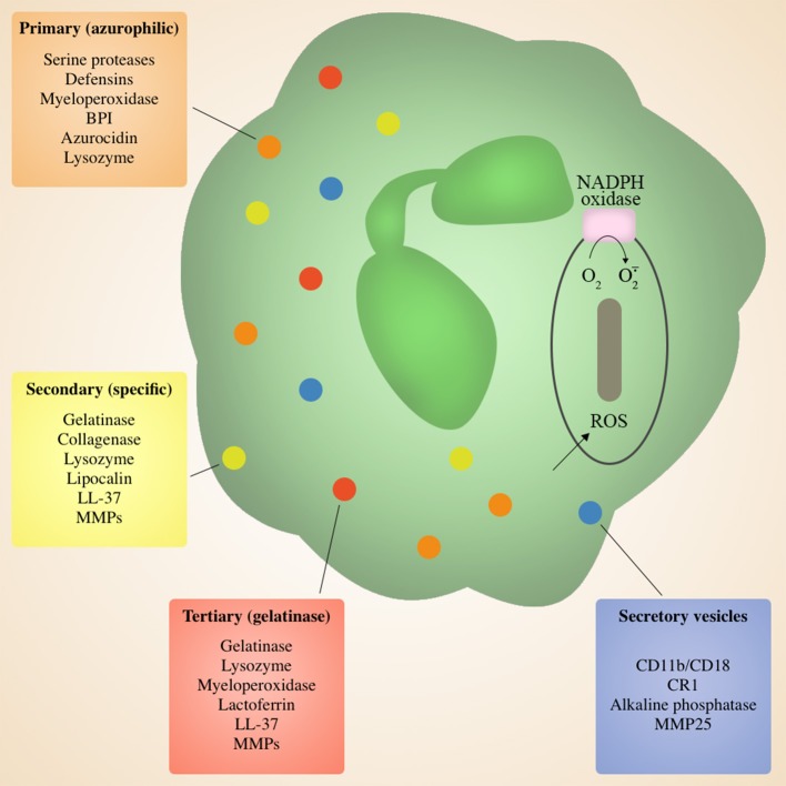
Pneumonia is a leading cause of death from infection in the United States and across the globe. During pulmonary infection, clear resolution of host inflammatory responses occurs in the absence of appreciable lung damage. Neutrophils are the first wave of leukocytes to arrive in the lung upon infection. After activation, neutrophils traffic from the vasculature via transendothelial migration through the lung interstitium and into the alveolar space. Successful pulmonary immunity requires neutrophil-mediated killing of invading pathogens by phagocytosis and release of a myriad of antimicrobial molecules, followed by resolution of inflammation, neutrophil apoptosis, and clearing of dead or dying neutrophils by macrophages. In addition to their antimicrobial role, it is becoming clear that neutrophils are also important modulators of innate and adaptive immune responses, primarily through the release of cytokines and recruitment of additional waves of neutrophils into the airways. Though typically essential to combating severe pneumonia, neutrophil influx into the airways is a double-edged sword: Overzealous neutrophil activation can cause severe tissue damage as a result of the release of toxic agents including proteases, cationic polypeptides, cytokines, and reactive oxygen species (ROS) aimed at killing invading microbes. In extreme cases, the damage caused by neutrophils and other innate immune mediators become the primary source of morbidity and mortality. Here, we review the complex role of neutrophils during severe pneumonia by highlighting specific molecules and processes that contribute to pulmonary immunity, but can also drive progression of severe disease. Depending on the identity of the infectious agent, enhancing or suppressing neutrophil-mediated responses may be key to effectively treating severe and typically lethal pneumonia.
Keywords: neutrophils; pneumonia; pulmonary damage; pulmonary immunity; pulmonary infection.
Figures
References
-
- Auten R. L., Mason S. N., Tanaka D. T., Welty-Wolf K., Whorton M. H. (2001). Anti-neutrophil chemokine preserves alveolar development in hyperoxia-exposed newborn rats. Am. J. Physiol. Lung Cell Mol. Physiol. 281, L336–L344. - PubMed
Publication types
MeSH terms
Substances
Grants and funding
LinkOut - more resources
Full Text Sources
Other Literature Sources
Medical