. 2017 Apr 27;5(4):2325967117702452.
doi: 10.1177/2325967117702452. eCollection 2017 Apr.

Early Osteoarthritis After Untreated Anterior Meniscal Root Tears: An In Vivo Animal Study

Affiliations

Early Osteoarthritis After Untreated Anterior Meniscal Root Tears: An In Vivo Animal Study

Brett D Steineman et al. Orthop J Sports Med. .

Abstract

Background: Meniscal root tears cause menisci and their insertions to inadequately distribute loads and potentially leave underlying articular cartilage unprotected. Untreated meniscal root tears are becoming increasingly recognized to induce joint degradation; however, little information is known about anterior meniscal root tears and how they affect joint tissue.

Purpose: To observe the early degenerative changes within the synovial fluid, menisci, tibial articular cartilage, and subchondral bone after arthroscopic creation of untreated anterior meniscal root tears.

Study design: Controlled laboratory study.

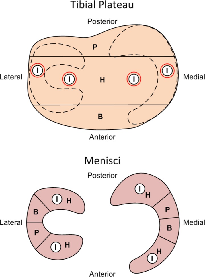
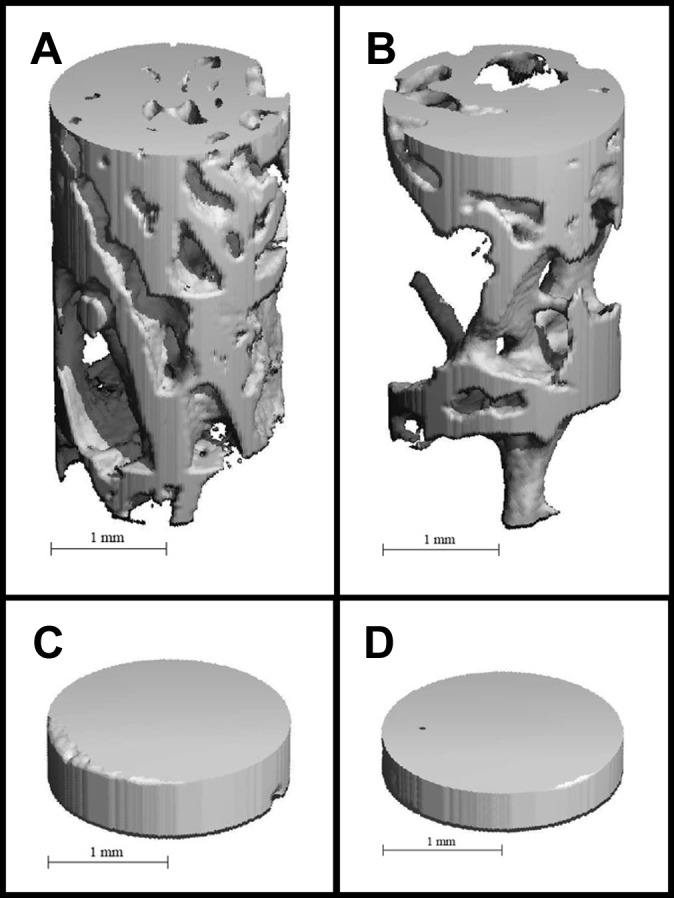
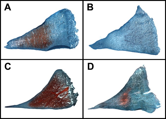
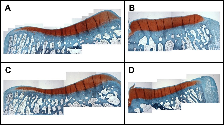
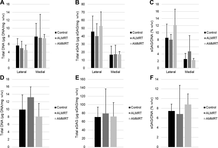
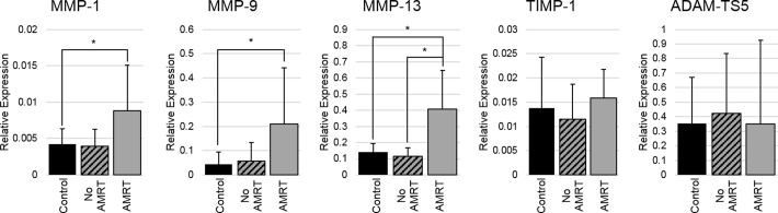
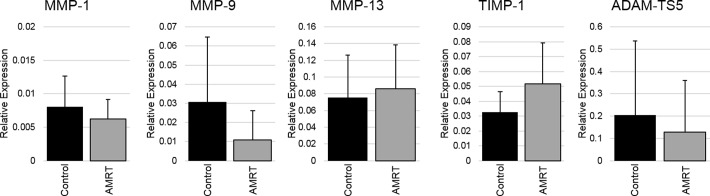
Methods: Anterolateral meniscal root tears were created in 1 knee joint of 5 adult Flemish Giant rabbits, and anteromedial meniscal root tears were created in 4 additional rabbits. The contralateral limbs were used as nonoperated controls. The animals were euthanized at 8 weeks postoperatively; synovial fluid was aspirated, and tissue samples of menisci and tibial articular cartilage were collected and processed for multiple analyses to detect signs of early degeneration.

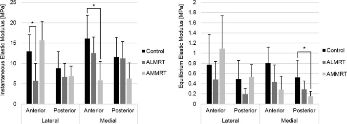
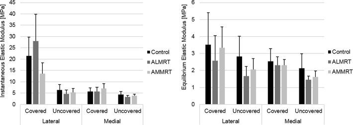
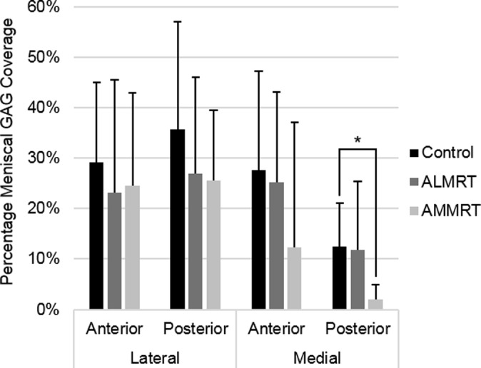
Results: Significant changes were found within the synovial fluid, meniscal tissue, and tibial subchondral bone of the knees with anterior meniscal root tears when compared with controls. There were no significant changes identified in the tibial articular cartilage when comparing the tear groups with controls.

Conclusion: This study demonstrated early degenerative changes within the synovial fluid, menisci, and tibial subchondral bone when leaving anterior meniscal root tears untreated for 8 weeks. The results suggest that meniscal tissue presents measurable, degenerative changes prior to changes within the articular cartilage after anterior meniscal root tears. Anterior destabilization of the meniscus arthroscopically may lead to measurable degenerative changes and be useful for future in vivo natural history and animal repair studies.

Clinical relevance: The present study is the first to investigate various tissue changes after anterior meniscal root tears of both the medial and lateral menisci. The results from this study suggest that degenerative changes occur within the synovial fluid, meniscus, and tibial subchondral bone prior to any measurable changes to the tibial articular cartilage. Further studies should expand on this study to evaluate how these components continue to progress when left untreated for long periods.

Keywords: animal model; anterior meniscus roots; meniscal root tear; meniscus; osteoarthritis.


Conflict of interest statement

One or more of the authors has declared the following potential conflict of interest or source of funding: R.F.L. receives royalties from Arthrex, Ossur, and Smith & Nephew; is a paid consultant for Arthrex, Ossur, and Smith & Nephew; and receives research support from Arthrex, Smith & Nephew, Ossur, and Linvatec. Funding for this project was provided by the Steadman Philippon Research Institute in Vail, Colorado.

Figures

Figure 1.
Figure 1.
A dissected, proximal tibia with menisci intact for visualization of the rabbit knee anatomy from a left, control limb.
Figure 2.
Figure 2.
Tissue locations allocated for different analyses on the menisci and tibial plateaus including: indentation-relaxation (I), histology (H), biochemical assays (B), and quantitative reverse transcription polymerase chain reaction (qRT-PCR) (P).
Figure 3.
Figure 3.
Instantaneous and equilibrium elastic moduli of meniscus regions (mean with standard deviation). *Statistically significant (P < .05). ALMRT, anterolateral meniscal root tear; AMMRT, anteromedial meniscal root tear.
Figure 4.
Figure 4.
Instantaneous and equilibrium elastic moduli of tibial articular cartilage regions (mean with standard deviation). *Statistically significant (P < .05). ALMRT, anterolateral meniscal root tear; AMMRT, anteromedial meniscal root tear.
Figure 5.
Figure 5.
Volume renderings of trabecular and subchondral bone volumes of interest (VOIs) in control and anterior meniscal root tear limbs. Examples are provided for trabecular bone VOI from (A) the lateral uncovered region in the control limb, (B) trabecular bone VOI from the lateral uncovered region in the anteromedial meniscal root tear limb, (C) subchondral bone VOI from the medial uncovered region in the control limb, and (D) subchondral bone VOI from the medial uncovered region in the anterolateral meniscal root tear limb.
Figure 6.
Figure 6.
Histological staining results of meniscal glycosaminoglycan (GAG) coverage (mean with standard deviation). *Statistically significant (P < .05). ALMRT, anterolateral meniscal root tear; AMMRT, anteromedial meniscal root tear.
Figure 7.
Figure 7.
Glycosaminoglycan staining in anterior meniscal regions for control and both anterior meniscal root tear limbs. Examples provided are (A) the posterior region of the medial meniscus in a control limb, (B) the posterior region of the medial meniscus in an anteromedial meniscal root tear limb, (C) the anterior region of the lateral meniscus in a control limb, and (D) the anterior region of the lateral meniscus in an anterolateral meniscal root tear limb.
Figure 8.
Figure 8.
Examples of glycosaminoglycan staining in the tibial articular cartilage for a control limb in the (A) lateral and (B) medial hemijoint as well as an anterolateral meniscal root tear limb in the (C) lateral and (D) medial hemijoint.
Figure 9.
Figure 9.
Total DNA content with respect to wet weight, total glycosaminoglycan (GAG) content with regard to wet weight, and total GAG with regard to total DNA content for menisci (A, B, and C, respectively) and tibial articular cartilage (D, E, and F, respectively). ALMRT, anterolateral meniscal root tear; AMMRT, anteromedial meniscal root tear.
Figure 10.
Figure 10.
Relative gene expressions for meniscus samples (mean with standard deviation). Anterior meniscal root tears (AMRT) of medial and lateral insertions were pooled for gene expression analysis. *Statistically significant (P < .05). ADAM-TS5, aggrecanase-2; MMP, matrix metalloproteinase; TIMP-1, tissue inhibitor of metalloproteinase-1.
Figure 11.
Figure 11.
Relative gene expressions for tibial articular cartilage samples (mean with standard deviation). Anterior meniscal root tears (AMRT) of medial and lateral insertions were pooled for gene expression analysis. *Statistically significant (P < .05). ADAM-TS5, aggrecanase-2; MMP, matrix metalloproteinase; TIMP-1, tissue inhibitor of metalloproteinase-1.

References

    1. Ahmed AM, Burke DL. In-vitro measurement of static pressure distribution in synovial joints—part I: tibial surface of the knee. J Biomech Eng. 1983;105:216–225. - PubMed
    1. Allaire R, Muriuki M, Gilbertson L, Harner CD. Biomechanical consequences of a tear of the posterior root of the medial meniscus. J Bone Joint Surg Am. 2008;90:1922–1931. - PubMed
    1. Allen AA, Caldwell GL, Fu FH. Anatomy and biomechanics of the meniscus. Oper Tech Orthop. 1995;5:2–9.
    1. Arunakul M, Tochigi Y, Goetz JE, et al. Replication of chronic abnormal cartilage loading by medial meniscus destabilization for modeling osteoarthritis in the rabbit knee in vivo. J Orthop Res. 2013;31:1555–1560. - PMC - PubMed
    1. Aspden RM, Yarker YE, Hukins DW. Collagen orientations in the meniscus of the knee joint. J Anat. 1985;140(pt 3):371–380. - PMC - PubMed

LinkOut - more resources