. 2017 May 10;3(5):e1602670.
doi: 10.1126/sciadv.1602670. eCollection 2017 May.

Cryo-EM structure of human adenovirus D26 reveals the conservation of structural organization among human adenoviruses

Affiliations

Cryo-EM structure of human adenovirus D26 reveals the conservation of structural organization among human adenoviruses

Xiaodi Yu et al. Sci Adv. .

Abstract

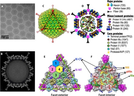
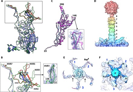
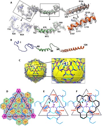
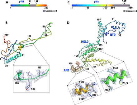
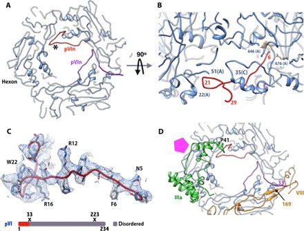
Human adenoviruses (HAdVs) cause acute respiratory, ocular, and gastroenteric diseases and are also frequently used as gene and vaccine delivery vectors. Unlike the archetype human adenovirus C5 (HAdV-C5), human adenovirus D26 (HAdV-D26) belongs to species-D HAdVs, which target different cellular receptors, and is differentially recognized by immune surveillance mechanisms. HAdV-D26 is being championed as a lower seroprevalent vaccine and oncolytic vector in preclinical and human clinical studies. To understand the molecular basis for their distinct biological properties and independently validate the structures of minor proteins, we determined the first structure of species-D HAdV at 3.7 Å resolution by cryo-electron microscopy. All the hexon hypervariable regions (HVRs), including HVR1, have been identified and exhibit a distinct organization compared to those of HAdV-C5. Despite the differences in the arrangement of helices in the coiled-coil structures, protein IX molecules form a continuous hexagonal network on the capsid exterior. In addition to the structurally conserved region (3 to 300) of IIIa, we identified an extra helical domain comprising residues 314 to 390 that further stabilizes the vertex region. Multiple (two to three) copies of the cleaved amino-terminal fragment of protein VI (pVIn) are observed in each hexon cavity, suggesting that there could be ≥480 copies of VI present in HAdV-D26. In addition, a localized asymmetric reconstruction of the vertex region provides new details of the three-pronged "claw hold" of the trimeric fiber and its interactions with the penton base. These observations resolve the previous conflicting assignments of the minor proteins and suggest the likely conservation of their organization across different HAdVs.

Keywords: Ad26; Cement proteins; Cryo-electron microscopy; HAdV-D26; Human adenovirus D26; Major capsid proteins; Minor capsid proteins; Structure.


Figures

Fig. 1
Fig. 1. Overall structure and organization of the major and minor capsid proteins in HAdV-D26.
(A) Representative micrograph of frozen-hydrated HAdV-D26 virions (defocus, 1.16 μm) (B) Slice through the center of HAdV-D26 particle showing the cross section of densities of the MCPs and closely associated minor proteins. The shaft repeats and knob domain of the fiber can be seen, and the packaged double-stranded DNA in the middle is disordered. (C) Radially color-coded surface representation of the cryo-EM reconstruction of HAdV-D26, a view down the icosahedral threefold axis. One icosahedral facet is identified by a black triangle. (D) Schematic representation of AdV capsid showing the location and organization of various proteins in the AdV capsid. A list of symbols and the proteins they represent is shown on the right. The copy numbers of the capsid proteins are indicated in parentheses, and “?” indicates the uncertainty in the copy numbers. (E) Exterior view of a triangular facet. The locations of the structurally distinct hexon-1 to hexon-4 are identified by differently colored hexagons, surface representations of the hexons (hexon-1 to hexon-4) are shown in four different colors (light blue, pink, green, and khaki, respectively), and the PB is shown in magenta. The green triangle represents an icosahedral facet and is equivalent to the black triangle in (C). The hexons shown in gray belong to neighboring facets. The locations of the NT triskelions and CT coiled coils (four-helical bundle; 4-HLXB) of protein IX are labeled. (F) Interior view of the facet showing the location of the minor proteins relative to each other and to the MCPs. Surface representations of the proteins IIIa, VIII-U, VIII-V, and pVIn are shown in dark green, orange, yellow, and red, respectively. With the exception of pVIn (red), multiple copies of the same-colored proteins are related to each other by the icosahedral symmetry.
Fig. 2
Fig. 2. Structures of MCPs in HAdV-D26.
(A) Superposition of the hexon subunits from HAdV-D26 in green and HAdV-C5 [Protein Data Bank (PDB) ID: 3IYN] in blue. The regions of the HAdV-C5 hexon, which differ from those of the HAdV-D26 by RMSDs >2.0 Å, are shown in red, and most of these differences occur in the HVRs. (B) Closeup view of the HVRs (some of them are labeled). The inset shows the EM density for HVR1. (C) Superposition of the PB subunits of HAdV-D26 (magenta) and HAdV-C5 (light blue; PDB ID: 3IYN). The inset shows a closeup view of the variable β34 loop with a six–amino acid insertion in the density. (D) Color-coded surface representation of the fiber density derived from the LAR that highlights the fiber shaft repeats with the receptor binding knob domain located at the top and the PB at the bottom. The eight fiber shaft repeats are labeled. (E) Cutaway top view of the extracted fiber density highlighting the elbow-shaped structures displayed by the fiber NT tails (FNTs). The three fiber tails of the “fiber claw” that are more strongly connected to the central fiber density are labeled as 1, 2, and 3. Arrows identify the locations of the breaks in the density of the remaining two FNTs at this contour level. The backbone trace of the FNT model (residues 2 to 20) derived from the high-resolution HAdV-D26 structure is shown inside the elbow-shaped densities (see also fig. S7). The Asp9 residue is located at the elbow bend. (F) Same view as in (E), showing the elbow-shaped FNTs (dark blue) hooking the base of the protrusion that contains the RGD loop, identified by asterisks (*). Numbers 1, 2, and 3 identify the three FNTs of the fiber claw, and the arrows indicate the break in the density of the remaining two FNTs.
Fig. 3
Fig. 3. Structure and organization of the protein IX molecules in HAdV-D26.
(A) A full-length molecule of IX ordered is shown in the corresponding EM density. The closeup views of different regions of the IX molecule in the EM density are shown below. (B) Ribbon diagram showing the secondary structure of the IX polypeptide with a 5-turn linker α helix in the middle and an 11-turn long α helix at the C terminus. (C) Continuous hexagonal network of IX molecules shown in the background of yellow icosahedron. A closeup view of the protein IX network is shown on the right. The four structurally distinct IX polypeptides (P, Q, R, and S) are shown in different colors (dark blue, light blue, cyan, and purple, respectively), and the same-colored copies are related by the icosahedral symmetry. Numbers 1 to 4 identify the locations of the unique hexons. The full-length molecule (R) ordered is shown in cyan, and the IX molecule (P) that contributes an antiparallel helix is shown in blue. Missing connections between the triskelions and coiled coils in P, Q, and S copies are identified by gray sticks. The triskelions at the icosahedral threefold axes are formed by purple IX molecules (S), whereas those at the local threefold axes are formed by three differently colored IX molecules (P, Q, and R). The four-helical bundle (4-HLXB) structures are formed by the CT helices of four structurally distinct IX-molecules that come from four different triskelions. The CT helices of the three IX molecules (Q, R, and S) are arranged in parallel and come from the same facet, whereas the fourth one (P), which joins in an antiparallel orientation, comes from the neighboring facet; this arrangement is similar to that of HAdV-C5. Asterisks (*) identify some of the 4-HLXBs. (D) Schematic diagram showing the hexagonal network of IX molecules shown as lines/sticks that interlace the hexons, which are represented as hexagons, whereas the PBs are shown as pentagons. The subunits (for example, A, B, and C) that constitute the individual hexons are labeled along with the locations of the V1 and V2 barrels of the individual hexon subunits. The centers of IX triskelions interact with the V2 domains of hexon subunits, whereas the 4-HLXB is located at the interface formed by V1 and V2 domains from two different subunits (K and L) of the hexon-4 capsomer on one side and their local twofold-related counterparts from D and E subunits of hexon-2 that belongs to the adjacent facet. The black triangle represents the boundary of the icosahedral facet. (E) Simplified schematic showing the path of the hexagonal network formed by protein IX molecules within a facet. The labels T and 4-HLXB identify the locations of the triskelions and coiled coils, respectively. The 4-HLXBs are encircled by red ovals. (F) Schematic identifying the locations of the triskelions and 4-HLXB with respect to the GON substructure, whose boundary is identified by the black lines overlaid on top of the protein IX network shown in (E). The 4-HLXBs are identified by red ovals.
Fig. 4
Fig. 4. Structures of the minor proteins VIII and IIIa.
(A) Schematic diagram showing the structurally ordered (colored)/disordered (gray) regions and the locations of proteolytic cleavage sites (X) in the precursor of VIII. (B) The structure of VIII (U) is elongated and has a shape of an obtuse triangle, with a length of ~100 Å and a width (height) of 30 Å. It is composed of two chains, comprising residues 1 to 110 and 158 to 227, which are the result of the proteolytic processing (at residues 111 and 157) by the AVP. The 47-residue middle fragment is disordered. The inset shows the representative electron density in the boxed region. (C) Schematic diagram showing the structurally ordered (colored)/disordered (gray) regions and the location of proteolytic cleavage site (X) in the precursor of IIIa. (D) The structure of the ordered regions of IIIa (amino acids 3 to 390) is mostly composed of α helices and spans 130 Å along the longest dimension and 50 Å wide. The structure of IIIa can be divided into three domains: the NT domain (NTD), the middle domain (MDLD), and the appendage domain (APD). Insets show representative EM densities in the MDLD and the APD. Residues 302 to 313 and 391 to 560 are disordered.
Fig. 5
Fig. 5. Structure and interactions of the cleaved NT fragment of VI (pVIn).
(A) Elongated structure and locations of two copies of pVIn bound to PPH. Only two of the three equivalent binding sites on PPH are occupied. (B) Side view showing the structure and interactions of one of the pVIn molecules. The N terminus of pVIn reaches deep into the hexon cavity near residues 646, 676, and 773, whereas residues 15 to 29 of pVIn interact with residues 48 to 84 of the A subunit and residues 29 to 35 of the C subunit at the entrance of the hexon cavity. (C) Top: Representative EM density for one of the pVIn molecules bound to PPH in (A) (in red; identified by the asterisk). Bottom: Schematic diagram of the structurally ordered (colored)/disordered (gray) regions and the locations of proteolytic cleavage sites (X) in the precursor of VI. (D) Interactions of pVIn with other minor proteins. Residues 25 to 29 of the above pVIn (red) interact with amino acids 42 to 48 of IIIa, whereas the CT residues (amino acids 30 to 33) of the second copy of pVIn (purple) form an antiparallel strand with residues 166 to 169 of VIII (U) (yellow). The pentagon (magenta) identifies the location of the PB.

References

    1. Davison A. J., Benkő M., Harrach B., Genetic content and evolution of adenoviruses. J. Gen. Virol. 84, 2895–2908 (2003). - PubMed
    1. Rux J. J., Burnett R. M., Type-specific epitope locations revealed by X-ray crystallographic study of adenovirus type 5 hexon. Mol. Ther. 1, 18–30 (2000). - PubMed
    1. Abbink P., Lemckert A. A. C., Ewald B. A., Lynch D. M., Denholtz M., Smits S., Holterman L., Damen I., Vogels R., Thorner A. R., O’Brien K. L., Carville A., Mansfield K. G., Goudsmit J., Havenga M. J. E., Barouch D. H., Comparative seroprevalence and immunogenicity of six rare serotype recombinant adenovirus vaccine vectors from subgroups B and D. J. Virol. 81, 4654–4663 (2007). - PMC - PubMed
    1. Bouard D., Alazard-Dany N., Cosset F.-L., Viral vectors: From virology to transgene expression. Br. J. Pharmacol. 157, 153–165 (2009). - PMC - PubMed
    1. Chen C. Y., Senac J. S., Weaver E. A., May S. M., Jelinek D. F., Greipp P., Witzig T., Barry M. A., Species D adenoviruses as oncolytics against B-cell cancers. Clin. Cancer Res. 17, 6712–6722 (2011). - PMC - PubMed

Substances

LinkOut - more resources