. 2017 May 10;3(5):e1700933.
doi: 10.1126/sciadv.1700933. eCollection 2017 May.

Structures of closed and open conformations of dimeric human ATM

Affiliations

Structures of closed and open conformations of dimeric human ATM

Domagoj Baretić et al. Sci Adv. .

Abstract

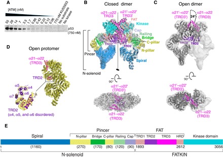
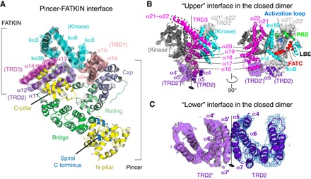
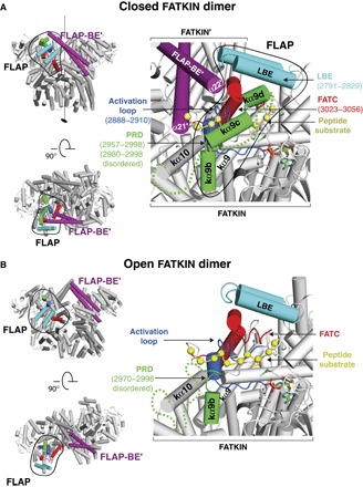
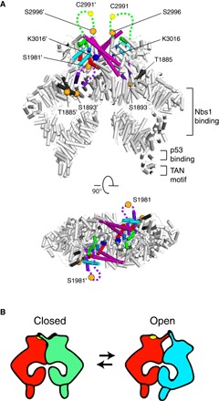
ATM (ataxia-telangiectasia mutated) is a phosphatidylinositol 3-kinase-related protein kinase (PIKK) best known for its role in DNA damage response. ATM also functions in oxidative stress response, insulin signaling, and neurogenesis. Our electron cryomicroscopy (cryo-EM) suggests that human ATM is in a dynamic equilibrium between closed and open dimers. In the closed state, the PIKK regulatory domain blocks the peptide substrate-binding site, suggesting that this conformation may represent an inactive or basally active enzyme. The active site is held in this closed conformation by interaction with a long helical hairpin in the TRD3 (tetratricopeptide repeats domain 3) domain of the symmetry-related molecule. The open dimer has two protomers with only a limited contact interface, and it lacks the intermolecular interactions that block the peptide-binding site in the closed dimer. This suggests that the open conformation may be more active. The ATM structure shows the detailed topology of the regulator-interacting N-terminal helical solenoid. The ATM conformational dynamics shown by the structures represent an important step in understanding the enzyme regulation.

Keywords: ATM; Ataxia telangiectasia mutated; DNA damage response; cryo-EM; kinase; structure.


Figures

Fig. 1
Fig. 1. Structures and activity of human ATM dimers.
(A) Basal catalytic activity of purified human ATM with full-length p53 substrate (visualized by 33P autoradiography). (B) Side and top views of the closed dimer are shown as ribbon diagrams colored by domains within EM densities at a resolution of 4.4 Å for the Pincer-FATKIN and 5.7 Å for the N-terminal Spiral. The twofold symmetry axis is shown as a black line. (C) Side and top views of the open dimer having two protomers at a resolution of 11.5 Å (fig. S5, 3D class C). One protomer and a second FATKIN were fit into the density. The right molecule in the open dimer is tilted about 24° with respect to the orientation it would have in the closed dimer. (D) The open protomer (from 3D classes C and D) model and the map at an overall resolution of 5.7 Å (the Pincer-FATKIN region at a resolution of 4.8 Å). The inset shows details of the TRD2 region, where much of it is disordered in the open protomer. Helices α4, α5, and α6 are not ordered in the open protomer, but for reference, they are shown in the inset, surrounded by a dashed yellow curve. (E) A bar diagram of human ATM (approximate numbers of residues in the N-solenoid domains are shown in parentheses).
Fig. 2
Fig. 2. Views of the Pincer-FATKIN interface and the dimer interface.
(A) A closeup view of the Pincer domains and their interactions with the FATKIN. (B) The bipartite interface between the two protomers in the closed dimer. The upper interface includes portions of the kinase domain and TRD3. (C) The lower interface, primarily including TRD2 and a portion of TRD3, shown as ribbons with the density of the closed dimer interface (purple and blue).
Fig. 3
Fig. 3. Open and closed ATM dimer conformations of the active site.
(A) The side view on the left shows the FATKIN arrangement in the closed dimer. The FLAP-BE′ (purple) from the symmetry-related molecule restricts the PRD to a conformation that blocks the substrate peptide from entering the active site (right panel). The location of ADP-F3Mg was modeled on the basis of the crystal structure of the mTOR FATKIN [Protein Data Bank (PDB) ID 4JSV]. The location of the substrate peptide (yellow spheres) was modeled by superimposing a peptide-bound Cdk2 [PDB ID 3QHW (50)] onto the ATM active site. (B) A model of the active site of the 4.8 Å resolution open protomer (right panel). The side view on the left is the arrangement of the two FATKIN moieties in the 11.5 Å resolution open dimer. The green dashed lines in each panel represent the disordered portion of the PRD.
Fig. 4
Fig. 4. A dynamic equilibrium of ATM dimers may be regulated by interactions with activators and substrates.
(A) A model of ATM highlighting known activating influences on ATM. The orange spheres denote locations of four sites of autophosphorylation (Ser1981 and Ser2996 are in disordered loops suggested by dashed lines). The FLAP-BE (purple), FATC (red), LBE (cyan), activation loop (light blue), and PRD (green) are highlighted. Cys2991, whose oxidation is associated with ATM activation, is marked by a yellow sphere in the disordered PRD loop (suggested by a dashed line). The Lys3016 acetylation site in the dimeric contact of the FATC is shown as a blue sphere. The approximate locations of the N-terminal TAN motif and the Nbs1 and p53 binding sites are indicated. (B) A schematic representation of the ATM dynamic equilibrium between open and closed dimers. Specific substrates or regulators could differentially recognize variations in the dimer interface.

References

    1. Shiloh Y., Ziv Y., The ATM protein kinase: Regulating the cellular response to genotoxic stress, and more. Nat. Rev. Mol. Cell. Biol. 14, 197–210 (2013). - PubMed
    1. Bakkenist C. J., Kastan M. B., DNA damage activates ATM through intermolecular autophosphorylation and dimer dissociation. Nature 421, 499–506 (2003). - PubMed
    1. Savitsky K., Sfez S., Tagle D. A., Ziv Y., Sartiel A., Collins F. S., Shiloh Y., Rotman G., The complete sequence of the coding region of the ATM gene reveals similarity to cell cycle regulators in different species. Hum. Mol. Genet. 4, 2025–2032 (1995). - PubMed
    1. Paull T. T., Mechanisms of ATM activation. Annu. Rev. Biochem. 84, 711–738 (2015). - PubMed
    1. Mavrou A., Tsangaris G. T., Roma E., Kolialexi A., The ATM gene and ataxia telangiectasia. Anticancer Res. 28, 401–405 (2008). - PubMed

MeSH terms

Substances