. 2017 Aug;134(2):255-269.
doi: 10.1007/s00401-017-1725-7. Epub 2017 May 15.

In-depth clinico-pathological examination of RNA foci in a large cohort of C9ORF72 expansion carriers

Affiliations

In-depth clinico-pathological examination of RNA foci in a large cohort of C9ORF72 expansion carriers

Mariely DeJesus-Hernandez et al. Acta Neuropathol. 2017 Aug.

Abstract

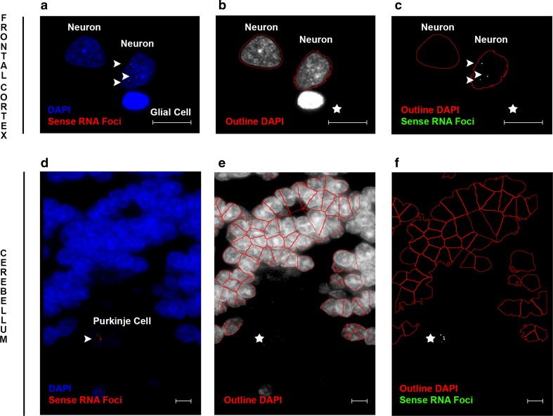
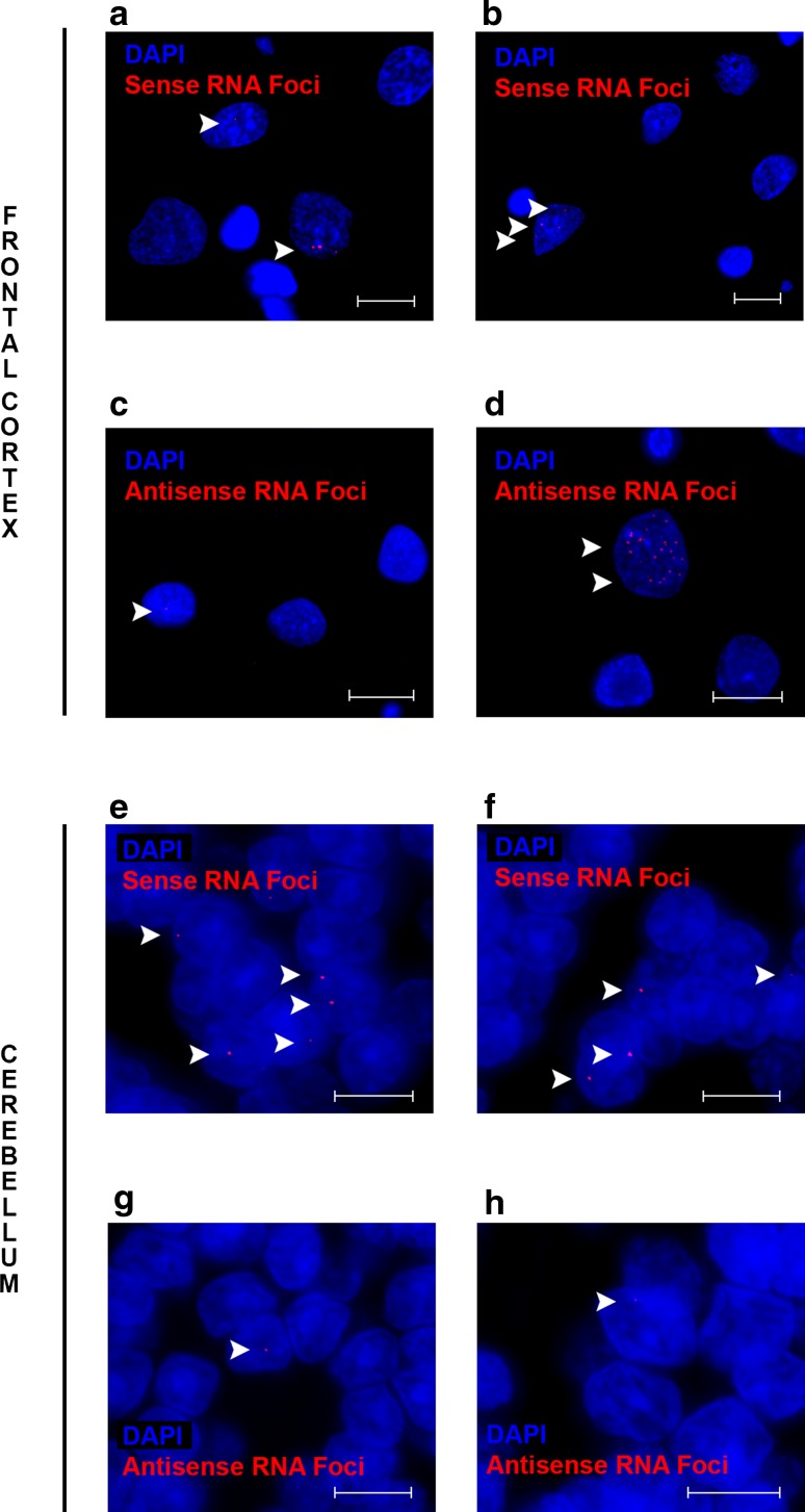
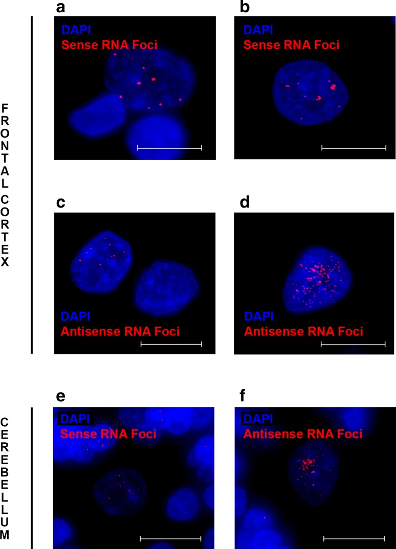
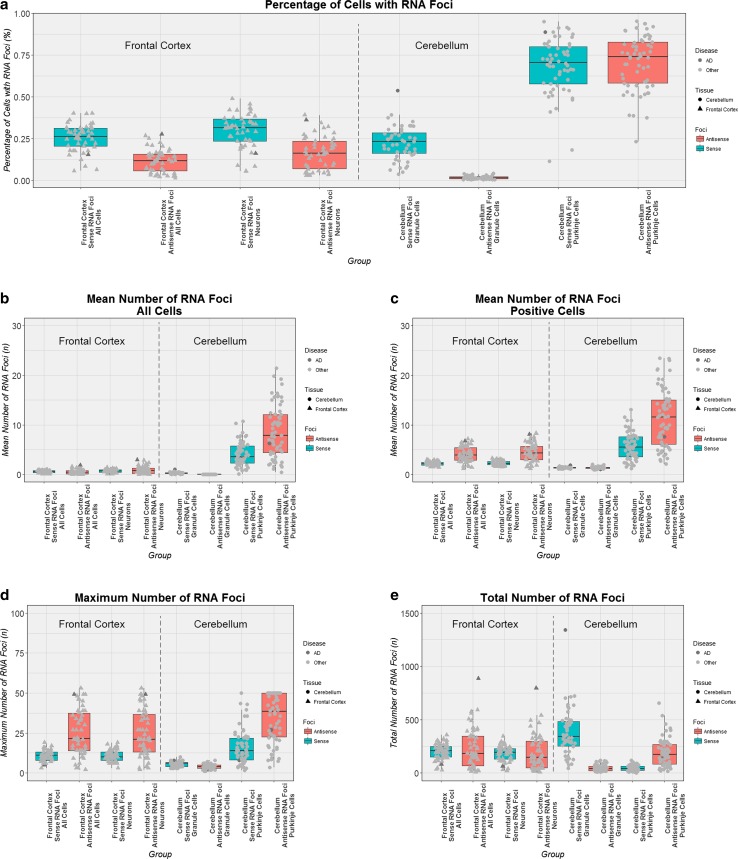
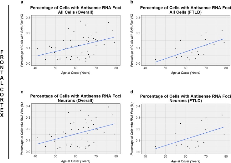
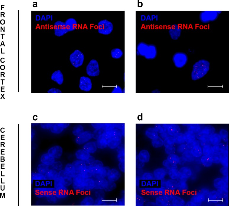
A growing body of evidence suggests that a loss of chromosome 9 open reading frame 72 (C9ORF72) expression, formation of dipeptide-repeat proteins, and generation of RNA foci contribute to disease pathogenesis in amyotrophic lateral sclerosis and frontotemporal dementia. Although the levels of C9ORF72 transcripts and dipeptide-repeat proteins have already been examined thoroughly, much remains unknown about the role of RNA foci in C9ORF72-linked diseases. As such, we performed a comprehensive RNA foci study in an extensive pathological cohort of C9ORF72 expansion carriers (n = 63). We evaluated two brain regions using a newly developed computer-automated pipeline allowing recognition of cell nuclei and RNA foci (sense and antisense) supplemented by manual counting. In the frontal cortex, the percentage of cells with sense or antisense RNA foci was 26 or 12%, respectively. In the cerebellum, 23% of granule cells contained sense RNA foci and 1% antisense RNA foci. Interestingly, the highest percentage of cells with RNA foci was observed in cerebellar Purkinje cells (~70%). In general, more cells contained sense RNA foci than antisense RNA foci; however, when antisense RNA foci were present, they were usually more abundant. We also observed that an increase in the percentage of cells with antisense RNA foci was associated with a delayed age at onset in the frontal cortex (r = 0.43, p = 0.003), whereas no other associations with clinico-pathological features were seen. Importantly, our large-scale study is the first to provide conclusive evidence that RNA foci are not the determining factor of the clinico-pathological variability observed in C9ORF72 expansion carriers and it emphasizes that the distribution of RNA foci does not follow the pattern of neurodegeneration, stressing the complex interplay between different aspects of C9ORF72-related diseases.

Keywords: Amyotrophic lateral sclerosis; C9ORF72; Frontotemporal dementia; Frontotemporal lobar degeneration; Motor neuron disease; RNA foci.


Conflict of interest statement

MDJ and RR hold a patent on methods to screen for the hexanucleotide repeat expansion in the C9ORF72 gene. All other authors declare that they have no conflicts of interest.

Figures

Fig. 1
Fig. 1
Examples of computer-automated recognition of specific cell types. Images are displayed to demonstrate the recognition of specific cell types in either the frontal cortex (top panel) or cerebellum (bottom panel). Cell nuclei (blue DAPI) and RNA foci (red RNA foci) are shown for images taken in the frontal cortex (a) and cerebellum (d). Our computer-automated pipelines are able to recognize neuronal nuclei in the frontal cortex (red outline nucleus), while ignoring glial nuclei (star; b). Additionally, they recognize cerebellar granule cells, without recognizing Purkinje cells that stain poorly with DAPI (star; e). RNA foci are shown in neuronal nuclei (green RNA foci; c); however, they are not being recognized in cerebellar Purkinje cells (star; f). RNA foci that might be difficult to see are highlighted by arrowheads. Scale bars 5 µm (top panel) or 2 µm (bottom panel)
Fig. 2
Fig. 2
Examples of frequently observed RNA foci patterns. Representative examples are shown of images with cell nuclei (blue DAPI) and RNA foci (red RNA foci) to demonstrate common patterns. In the frontal cortex, sense RNA foci are generally absent or a small number is seen (a, b). Antisense RNA foci are less frequently encountered, but if cells contain antisense RNA foci, then their numbers vary from just a few (c) to many (d). In the cerebellum, granule cells generally contain either no sense RNA foci or a relatively low number (e, f). Cerebellar antisense RNA foci are rare in granule cells, and if they are present, only one or two are detected (g, h). RNA foci that might be difficult to see are highlighted by arrowheads. Scale bars 5 µm (frontal cortex) or 2 µm (cerebellum)
Fig. 3
Fig. 3
Examples of cells with a large number of RNA foci. Occasionally, cells with abundant RNA foci are observed, both in the frontal cortex (ad) and cerebellum (e, f). Images are shown that display numerous RNA foci (red RNA foci) within a single nucleus (blue DAPI). Sense RNA foci are included (a, b, e) as well as antisense RNA foci (c, d, f). Please note the faint DAPI signal in Purkinje cells as compared to other cells (e, f). Scale bars 5 µm
Fig. 4
Fig. 4
Cell type- and tissue-specific differences in RNA foci measurements. Every box plot visualizes a different RNA foci measurement: the percentage of cells with RNA foci (a), the mean number of RNA foci in all cells (b), the mean number of RNA foci in cells with foci (c), the maximum number of RNA foci (d), and the total number of RNA foci (e). For each box plot, the median is represented by a solid black line, and each box spans the interquartile range (IQR; 25th percentile to 75th percentile). On the left side of all plots the frontal cortex is displayed, where each patient is represented by a solid triangle. On the right side the cerebellum is shown with solid circles representing patients. One expansion carrier with a primary pathological diagnosis of Alzheimer’s disease (AD) is highlighted in dark grey, whereas other patients are shown in light grey. Boxes that are turquoise denote sense RNA foci, whereas salmon boxes denote antisense RNA foci
Fig. 5
Fig. 5
Associations with age at onset. In the frontal cortex, associations with age at onset are shown for our overall cohort of C9ORF72 expansion carriers, either in all cells (a) or in neuronal cells (c). Additionally, associations are displayed for the subset of patients diagnosed with frontotemporal lobar degeneration (FTLD), again for all cells (b) or when enriching for neurons (d). The solid blue line denotes the linear regression line, while each individual is represented by a solid black circle
Fig. 6
Fig. 6
Examples of RNA foci in patient with Alzheimer’s disease (AD). Representative images are shown of a patient with primarily AD pathology who harbored numerous antisense RNA foci in the frontal cortex (a, b) as well as sense RNA foci in the cerebellum (c, d). Images contain cell nuclei (blue DAPI) and RNA foci (red RNA foci). Scale bars 5 µm (top panel) or 2 µm (bottom panel)
Fig. 7
Fig. 7
Transcript levels, dipeptide-repeat levels, and repeat length for patient with Alzheimer’s disease (AD). In the frontal cortex (turquoise boxes), the expression levels of all C9ORF72 expansion carriers are shown for C9ORF72 transcript variant 1 (a) as well as for intron 1b containing C9ORF72 transcripts (b). Similarly, poly(GP) dipeptide-repeat protein levels (c) and the length of the C9ORF72 expansion (d) are displayed of all C9ORF72 expansion carriers in the cerebellum (salmon boxes). For each box plot, the median is represented by a solid black line, and each box spans the interquartile range (IQR; 25th percentile to 75th percentile). A solid dark grey circle is used to denote an expansion carrier with a primary pathological diagnosis of AD; other patients are shown in light grey

References

    1. Almeida S, Gascon E, Tran H, Chou HJ, Gendron TF, Degroot S, Tapper AR, Sellier C, Charlet-Berguerand N, Karydas A, et al. Modeling key pathological features of frontotemporal dementia with C9ORF72 repeat expansion in iPSC-derived human neurons. Acta Neuropathol. 2013;126:385–399. doi: 10.1007/s00401-013-1149-y. - DOI - PMC - PubMed
    1. Ash PE, Bieniek KF, Gendron TF, Caulfield T, Lin WL, Dejesus-Hernandez M, van Blitterswijk MM, Jansen-West K, Paul JW, 3rd, Rademakers R, et al. Unconventional translation of C9ORF72 GGGGCC expansion generates insoluble polypeptides specific to c9FTD/ALS. Neuron. 2013;77:639–646. doi: 10.1016/j.neuron.2013.02.004. - DOI - PMC - PubMed
    1. Bieniek KF, Murray ME, Rutherford NJ, Castanedes-Casey M, DeJesus-Hernandez M, Liesinger AM, Baker MC, Boylan KB, Rademakers R, Dickson DW. Tau pathology in frontotemporal lobar degeneration with C9ORF72 hexanucleotide repeat expansion. Acta Neuropathol. 2013;125:289–302. doi: 10.1007/s00401-012-1048-7. - DOI - PMC - PubMed
    1. Bieniek KF, van Blitterswijk M, Baker MC, Petrucelli L, Rademakers R, Dickson DW. Expanded C9ORF72 hexanucleotide repeat in depressive pseudodementia. JAMA Neurol. 2014;71:775–781. doi: 10.1001/jamaneurol.2013.6368. - DOI - PMC - PubMed
    1. Cacace R, Van Cauwenberghe C, Bettens K, Gijselinck I, van der Zee J, Engelborghs S, Vandenbulcke M, Van Dongen J, Baumer V, Dillen L, et al. C9orf72 G4C2 repeat expansions in Alzheimer’s disease and mild cognitive impairment. Neurobiol Aging. 2013;34(1712):e1711–e1717. - PubMed

MeSH terms