. 2017 May 16:8:15205.
doi: 10.1038/ncomms15205.

Mutant KRAS promotes malignant pleural effusion formation

Affiliations

Mutant KRAS promotes malignant pleural effusion formation

Theodora Agalioti et al. Nat Commun. .

Abstract

Malignant pleural effusion (MPE) is the lethal consequence of various human cancers metastatic to the pleural cavity. However, the mechanisms responsible for the development of MPE are still obscure. Here we show that mutant KRAS is important for MPE induction in mice. Pleural disseminated, mutant KRAS bearing tumour cells upregulate and systemically release chemokine ligand 2 (CCL2) into the bloodstream to mobilize myeloid cells from the host bone marrow to the pleural space via the spleen. These cells promote MPE formation, as indicated by splenectomy and splenocyte restoration experiments. In addition, KRAS mutations are frequently detected in human MPE and cell lines isolated thereof, but are often lost during automated analyses, as indicated by manual versus automated examination of Sanger sequencing traces. Finally, the novel KRAS inhibitor deltarasin and a monoclonal antibody directed against CCL2 are equally effective against an experimental mouse model of MPE, a result that holds promise for future efficient therapies against the human condition.


Conflict of interest statement

L.A.S. is an employee of the company that produces the anti-CCL2 antibodies. The remaining authors declare no competing financial interests.

Figures
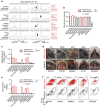
Figure 1
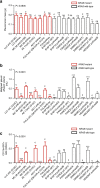
Figure 1. Selective induction of malignant pleural effusions by KRAS-mutant tumour cells.
Mutation status of and malignant pleural disease induction by twelve murine and human tumour cell lines after pleural delivery to appropriate recipient mice. (a) Kras cDNA Sanger sequencing traces of C57BL/6 mouse splenocytes (control) and of five C57BL/6 mouse tumour cell lines. Black arrows indicate heterozygous missense mutations in Kras codons 12 and 13. (b) Data summary of pleural tumor mass (n=53, 26, 19, 30, 19, 27, 20, 16, 14, 14, 14, and 15, respectively, for LLC, MC38, AE17, B16F10, PANO2, FULA, CULA, A549, LTP A549, SKMEL2, HT-29, and HEK293T cells). (c) Data summary of malignant pleural effusion (MPE) volume (n=53, 26, 19, 30, 19, 27, 20, 16, 14, 14, 14, and 15, respectively, for LLC, MC38, AE17, B16F10, PANO2, FULA, CULA, A549, LTP A549, SKMEL2, HT-29, and HEK293T cells). (d) Representative images of MPEs (dashed lines), pleural tumours (t), lungs (l), and hearts (h) imaged through the diaphragm. Scale bars, 1 cm. (e) Data summary of pleural CD11b+Gr1+ cells (n=5–16 animals/group were analysed). (f) Representative dotplots and gating strategy for the quantification of pleural CD11b+Gr1+ cells. Data are presented as mean±s.e.m. P, probability values for overall comparisons by one-way ANOVA. * and ***: P<0.05 and P<0.001 for the comparison between HEK293T cells and any other cell line (b) or for the comparison between any Kras-mutant and any Kras-wild-type cell line (c,e) by Bonferroni post-tests. WT, wild-type; LLC, C57BL/6 Lewis lung carcinoma; MC38, C57BL/6 colon adenocarcinoma; AE17, C57BL/6 malignant pleural mesothelioma; B16F10, C57BL/6 malignant skin melanoma; PANO2, C57BL/6 pancreatic adenocarcinoma; FULA, FVB urethane-induced lung adenocarcinoma; CULA, C57BL/6 urethane-induced lung adenocarcinoma; A549, human lung adenocarcinoma; LTP A549, long-term passaged A549 cells having lost the Y chromosome; SKMEL2, human malignant skin melanoma; HT-29, human colon adenocarcinoma; HEK293T, human embryonic kidney cells.
Figure 2
Figure 2. Mutant KRAS promotes malignant pleural effusion development.
Impact of shRNA-mediated Kras silencing on MPE competence of cell lines harboring mutant Kras, and of mutant KrasG12C overexpression in cell lines harboring wild-type KRAS. (a) Data summary of pleural tumor mass (n=14, 12, 11, 14, 11, 11, 9, 9, 11, 10, 13, 16, 12, 16, 9, 10, 9, and 9, respectively, for LLC shC, LLC shKras, MC38 shC, MC38 shKras, AE17 shC, AE17 shKras, FULA shC, FULA shKras, B16F10 pC, B16F10 pΔKras2A, B16F10 pΔKras2B, PANO2 pC, PANO2 pΔKras2A, PANO2 pΔKras2B, SKMEL2 pC, SKMEL2 pΔKras2B, HEK293T pC, and HEK293T pΔKras2B cells). (b) Data summary of MPE volume (n=14, 12, 11, 14, 11, 11, 9, 9, 11, 10, 13, 16, 12, 16, 9, 10, 9, and 9, respectively, for LLC shC, LLC shKras, MC38 shC, MC38 shKras, AE17 shC, AE17 shKras, FULA shC, FULA shKras, B16F10 pC, B16F10 pΔKras2A, B16F10 pΔKras2B, PANO2 pC, PANO2 pΔKras2A, PANO2 pΔKras2B, SKMEL2 pC, SKMEL2 pΔKras2B, HEK293T pC, and HEK293T pΔKras2B cells). (c) Data summary of pleural CD11b+Gr1+ cells (n=7–11/group were analysed). Data are presented as mean±s.e.m. P, probability values for overall comparisons by one-way ANOVA. ns, *, **, and ***: P>0.05, P<0.05, P<0.01, and P<0.001 for the comparison between the indicated cell line and the respective control (c) by Student's t-test or one-way ANOVA with Bonferroni post-tests, as appropriate. WT, wild-type; shC, random shRNA; shKras, anti-Kras-specific shRNA; pC, control (empty) overexpression vector; pΔKras2A and pΔKras2B, overexpression vectors encoding mutant mouse KrasG12C isoforms A and B, respectively; WT, wild-type; LLC, C57BL/6 Lewis lung carcinoma; MC38, C57BL/6 colon adenocarcinoma; AE17, C57BL/6 malignant pleural mesothelioma; B16F10, C57BL/6 malignant skin melanoma; PANO2, C57BL/6 pancreatic adenocarcinoma; FULA, FVB urethane-induced lung adenocarcinoma; SKMEL2, human malignant skin melanoma; HEK293T, human embryonic kidney cells.
Figure 3
Figure 3. Mutant KRAS signals via CCL2 to recruit splenic myeloid cells to malignant pleural effusions.
(a) Comparative transcriptome analysis of mouse tumour cell lines with defined Kras mutation status versus benign airway epithelial cells by microarray. Diagram depicting the analytic strategy employed to identify the transcriptional signature of mutant Kras comprised of 25 genes (top ten shown in table), among which Ccl2 ranked second. (b) Chemokine protein secretion by parental (white bars: cells stably expressing random shRNA or control overexpression vector) and Kras-modulated (red bars: cells stably expressing anti-Kras-specific shRNA; green bars: cells stably expressing vector encoding mutant mouse KrasG12C isoform B) murine cell lines by ELISA showing transcriptional regulation of CCL2, but not of CXCL1 and CXCL2, by mutant Kras (n=5–7/group). (c) Data summaries of malignant pleural effusion (MPE) volume (top; LLC: n=9/group; MC38: n=14–15/group; PANO2 pΔKras2A: n=8–18/group) and pleural CD11b+Gr1+ cells (bottom; LLC: n=9/group; MC38: n=14–15/group; PANO2 pΔKras2A: n=5/group) of Ccr2−/− and C57BL/6 control mice after intrapleural injection of three different tumour cell lines. pΔKras2A, vector encoding mouse KrasG12C isoform A. (d) Representative bioluminescent images of chimeric C57BL/6 mouse transplanted with bioluminescent bone marrow from CAG.Luc.eGFP donor before and after splenectomy performed at day 13 after intrapleural MC38 cells. Scale bars, 1 cm. (e) Data summaries of MPE volume (top; n=9/group) and pleural CD11b+Gr1+ cells (bottom; n=9/group) of C57BL/6 mice pretreated with sham surgery or splenectomy followed by intrapleural injection of MC38 cells, or PANO2 cells expressing pΔKras2A or pΔKras2B two weeks later. Data are presented as mean±s.d. ns, *, **, and ***: P>0.05, P<0.05, P<0.01, and P<0.001 for comparison with parental lines (b), between the two mouse strains (c), or between different surgeries (e) by Student's t-test.
Figure 4
Figure 4. KRAS mutations in human malignant pleural effusions.
(ac) Sanger sequencing results of human malignant pleural effusions (MPE) caused by metastatic lung adenocarcinomas from Institution 1. (a) Partial sequence of Homo Sapiens KRAS isoform b transcript showing start codon (green box) and missense mutations identified (grey boxes and callouts). Red and blue fonts indicate, respectively, known pathogenic mutations and mutations of unknown significance based on COSMIC. (b) Partial Sanger-sequencing traces from two patients showing corresponding sequences of patient with wild-type KRAS alleles and of another with four different KRAS mutations. Arrows indicate missense mutations of pathogenic (red) and unknown (blue) significance based on COSMIC, as well as nonsense mutations (grey). Note that mutant KRAS traces hide under wild-type traces superimposed by wild-type KRAS alleles and/or by RNA from tumour or MPE-infiltrating benign somatic cells. Importantly, some mutations were not detected by the analysis software (see letters above mutant trace). Note also multiple mutations in the same patient suggesting a possible multiclonal origin of this MPE. (ce) Patient-derived MPE cell line isolation from eight patients from Institution 1 that were initially tested KRAS wild-type. (c) Arrow shows focal clonal expansion of cultured MPE cells that gave rise to cell line PB-183. Scale bars, 50 μm. (d) PB-183-induced tumour in NOD/SCID mouse four weeks after subcutaneous injection of a million cells (n=5). Scale bar, 1 cm. (e) Partial Sanger-sequencing traces of KRAS cDNA from the initial MPE cells and from two MPE-derived cell lines indicate KRAS mutations (red arrows and fonts) that were not identified in the initial samples. Note that even in MPE cell lines mutant KRAS traces hide under wild-types traces superimposed by wild-type alleles. Again, the mutation was not detected by the software (see letters above mutant trace).
Figure 5
Figure 5. Mutant KRAS-mediated malignant pleural effusions are actionable.
(a) C57BL/6 mice received pleural MC38 cells (ΔKrasG13R), were allowed seven days for pleural tumour development, and were randomized to daily intraperitoneal saline (100 μl) or deltarasin (15 mg kg−1) treatments. Shown are data summaries of malignant pleural effusion (MPE) volume and CD11b+r1+ cells (both n=8/group), representative images of pleural effusions (dashed lines) and tumours (t), and representative dotplots of CD11b+Gr1+ cells (polygon gates) at day 13 post-MC38 cells. Scale bars, 1 cm. (b) MC38 cells were treated in vitro with saline or deltarasin (15 μgml−1). Shown is CCL2 secreted at 24 h (n=5/group). (c) NOD/SCID mice received pleural LTP A549 cells (ΔKRASG12S), were allowed 14 days for pleural tumour development, and were randomized to daily intraperitoneal saline (100 μl) or deltarasin (15 mg kg−1) treatments. Shown is data summary of MPE volume at day 30 post-tumour cells. (d) C57BL/6 mice received pleural MC38 cells followed by a single intrapleural injection of liposomes containing 1% DMSO or 15 mg kg−1 deltarasin in 1% DMSO at day 7 post-tumour cells. Shown are representative images of pleural effusions (dashed lines) and tumours (t), and data summaries of MPE volume (n=15–16/group) and CD11b+Gr1+ cells (n=9/group) at day 13 post-MC38 cells. Scale bars, 1 cm. (e) C57BL/6 mice received pleural PANO2 cells stably expressing mutant Kras vectors (pΔKras2A or pΔKras2B), were allowed 4 or 14 days, respectively, for pleural tumour development and were then randomized to intraperitoneal treatment with daily saline plus IgG2a antibody every three days (50 mg kg−1 in 100 μl saline), daily deltarasin (15 mg kg−1 in 100 μl saline), or anti-CCL2 antibody every three days (50 mg kg−1 in 100 μl saline). Shown are data summaries of MPE volume (n 27, 10, and 20 mice/group, respectively) and CD11b+Gr1+ cells (n=24, 8, and 14/group, respectively) at day 14 post-tumour cells. Data are presented as mean±s.d. ns, *, **, and ***: P>0.05, P<0.05, P<0.01, and P<0.001 for the indicated comparisons by Student's t-test (a-d) or one-way ANOVA with Bonferroni post-tests (e).

References

    1. Davies H. E. et al.. Effect of an indwelling pleural catheter versus chest tube and talc pleurodesis for relieving dyspnea in patients with malignant pleural effusion: the TIME2 randomized controlled trial. JAMA 307, 2383–2389 (2012). - PubMed
    1. Stathopoulos G. T. & Kalomenidis I. Malignant pleural effusion: tumor-host interactions unleashed. Am. J. Respir. Crit. Care Med. 186, 487–492 (2012). - PMC - PubMed
    1. Ryu J. S. et al.. Prognostic impact of minimal pleural effusion in non-small-cell lung cancer. J. Clin. Oncol. 32, 960–967 (2014). - PubMed
    1. Wu S. G. et al.. Survival of lung adenocarcinoma patients with malignant pleural effusion. Eur. Respir. J. 41, 1409–1418 (2013). - PubMed
    1. Stathopoulos G. T. et al.. A central role for tumor-derived monocyte chemoattractant protein-1 in malignant pleural effusion. J. Natl. Cancer Inst. 100, 1464–1476 (2008). - PubMed

Publication types

MeSH terms