. 2017 May 16:8:15287.
doi: 10.1038/ncomms15287.

Exosomes maintain cellular homeostasis by excreting harmful DNA from cells

Affiliations

Exosomes maintain cellular homeostasis by excreting harmful DNA from cells

Akiko Takahashi et al. Nat Commun. .

Erratum in

Abstract

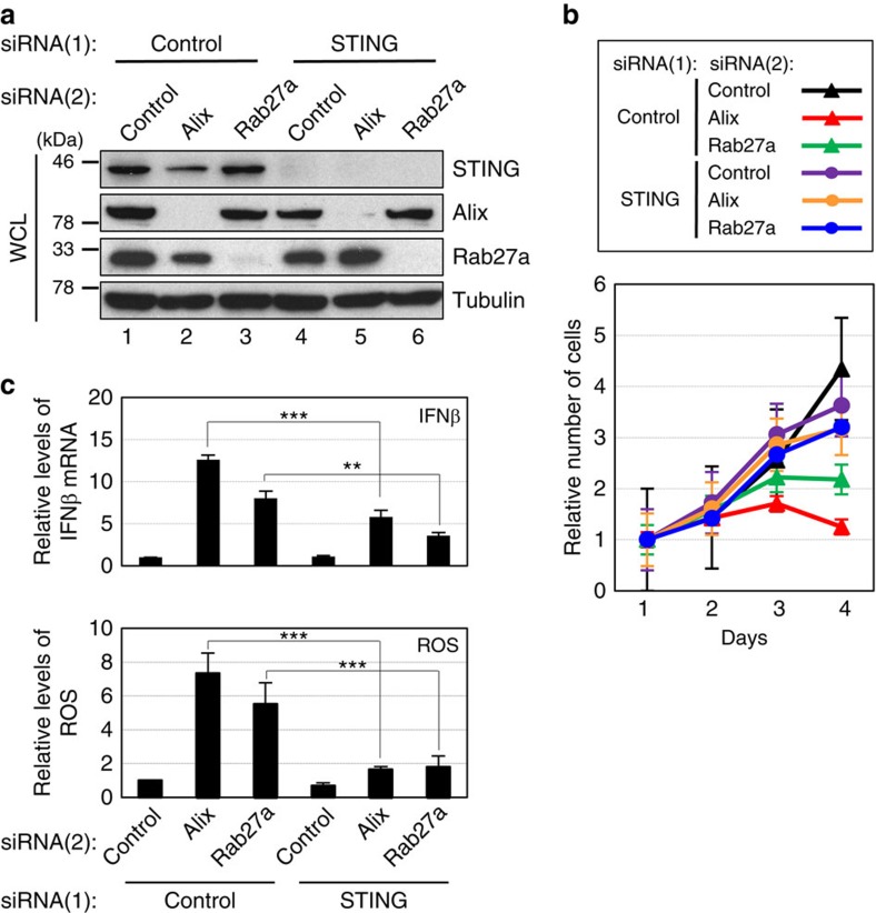
Emerging evidence is revealing that exosomes contribute to many aspects of physiology and disease through intercellular communication. However, the biological roles of exosome secretion in exosome-secreting cells have remained largely unexplored. Here we show that exosome secretion plays a crucial role in maintaining cellular homeostasis in exosome-secreting cells. The inhibition of exosome secretion results in the accumulation of nuclear DNA in the cytoplasm, thereby causing the activation of cytoplasmic DNA sensing machinery. This event provokes the innate immune response, leading to reactive oxygen species (ROS)-dependent DNA damage response and thus induce senescence-like cell-cycle arrest or apoptosis in normal human cells. These results, in conjunction with observations that exosomes contain various lengths of chromosomal DNA fragments, indicate that exosome secretion maintains cellular homeostasis by removing harmful cytoplasmic DNA from cells. Together, these findings enhance our understanding of exosome biology, and provide valuable new insights into the control of cellular homeostasis.


Conflict of interest statement

The authors declare no competing financial interests.

Figures

Figure 1
Figure 1. Inhibition of exosome secretion in senescent HDFs.
(a,b) Senescent TIG-3 cells induced by serial passage (a) or oncogenic Ras expression (b) were transfected with validated siRNA oligos indicated at the top of the panel for twice at 2 day intervals. These cells were then subjected to western blotting using antibodies shown right (WCL) or to exosome isolation followed by western blotting using antibodies against canonical exosome markers shown right (exosome) and NanoSight analysis (NTA) for quantitative measurement of isolated exosome particles. The representative data from three independent experiments are shown. Tubulin was used as a loading control. (cf) Senescent TIG-3 cells described in a,b were subjected to cell proliferation analysis (c,d) or to apoptosis analysis at day 4 (e,f). The representative data from three independent experiments are shown. For all graphs, error bars indicate mean±s.d. of triplicate measurements. (**P<0.01, ***P<0.001; one-way analysis of variance).
Figure 2
Figure 2. Inhibition of exosome secretion in pre-senescent HDFs.
(a) Pre-senescent TIG-3 cells were subjected to transfection with indicated siRNA oligos twice (at 2 day intervals). These cells were then subjected to western blotting using antibodies shown right (WCL) or to exosome isolation followed by western blotting using antibodies against canonical exosome markers shown right (exosome) and NanoSight analysis (NTA) for quantitative measurement of isolated exosome particles. The representative data from three independent experiments are shown. Tubulin was used as a loading control. (bd) Pre-senescent TIG-3 cells cultured under the conditions described in a were subjected to cell proliferation analysis (b), apoptosis analysis at day 4 (c) or to immunofluorescence staining for markers of DNA damage (γ-H2AX [red], phosphor-Ser/Thr ATM/ATR (pST/Q) substrate [green] and 4′,6-diamidino-2-phenylindole [blue]) (d). The representative data from three independent experiments are shown. The histograms indicate the percentage of nuclei that contain more than 3 foci positive for both γ-H2AX and pST/Q staining (d). At least 100 cells were scored per group (d). (e,f) Pre-senescent TIG-3 cells were infected with retrovirus encoding flag-tagged wild-type Alix or Rab27a protein containing a mutated siRNA cleavage site (lanes 3 and 4) or empty vector (lanes 1 and 2). After selection with puromycin, cells were transfected with indicated siRNA oligos and then subjected to western blotting using antibodies shown right, NanoSight analysis for quantitative measurement of isolated exosome particles, apoptosis analysis at day 4 or to immunofluorescence staining for markers of DNA damage. Tubulin was used as a loading control. The representative data from three independent experiments are shown. For all graphs, error bars indicate mean±s.d. of triplicate measurements. (**P<0.01. ***P<0.001; one-way analysis of variance).
Figure 3
Figure 3. Exosomes secretion prevents ATM/ATR-dependent DDR.
Pre-senescent TIG-3 cells were transfected with two different sets of validated siRNA oligos indicated at the top of the panel for twice at 2 day intervals. These cells were then subjected to western blotting using antibodies shown right (a) or to cell proliferation analysis (b). Tubulin was used as a loading control (a). The representative data from three independent experiments are shown. Error bars indicate mean±s.d. of triplicate measurements.
Figure 4
Figure 4. Exosomes contain chromosome fragments.
(a) transmission electron microscopy micrograph of MVE in pre-senescent TIG-3 cells following immuno-gold labelling for dsDNA. Gold particles are depicted as black dots. Right image shows a digitally zoomed area of exosome. (b,c) Exosomal DNA isolated from pre-senescent TIG-3 cells were subjected to size distribution analysis using Electrophoresis Bioanalyzer system (b) or to deep sequencing analysis (c). Genomic DNA of TIG-3 cells are also subjected to deep sequencing analysis, as control (c). The read count of each 500-kb bin was normalized to RPKM and corrected by the mappability (c). (d) Purified exosomes from pre-senescent TIG-3 cells were subjected to sucrose density-gradient separation followed by western blotting using antibodies shown right, NanoSight analysis (NTA) for quantitative measurement of isolated exosome particles and quantitative PCR analysis for detection of genomic DNA fragments (GRM7 and GPC6).
Figure 5
Figure 5. Overexpression of Dnase2a attenuated the effects of Alix or Rab27a knockdown in HDFs.
Pre-senescent TIG-3 cells were infected with retrovirus encoding flag-tagged Dnase2a (lanes 4–6) or empty vector (lanes 1–3). After selection with puromycin, cells were transfected with indicated siRNA oligos and then subjected to western blotting using antibodies shown right (a), NanoSight analysis (NTA) for quantitative measurement of isolated exosome particles and western blotting using antibodies against canonical exosome markers shown right (exosome) (b), isolation of cytoplasmic fraction followed by quantitative PCR (qPCR) analysis of chromosomal DNA (c), immunofluorescence staining for markers of DNA damage (γ-H2AX [red], pST/Q (green) and 4′,6-diamidino-2-phenylindole (blue)) (d), qPCR analysis of IFNβ gene expression (e), analysis of intracellular ROS levels (e) or to apoptosis analysis at day 4 (e). Tubulin was used as a loading control (a). The histograms indicate the percentage of nuclei that contain more than 3 foci positive for both γ-H2AX and pST/Q staining (d). At least 100 cells were scored per group (d). The representative data from three independent experiments are shown. For all graphs, error bars indicate mean±s.d. of triplicate measurements. (**P<0.01. ***P<0.001; one-way analysis of variance).
Figure 6
Figure 6. Reduction of ROS levels attenuated the effects of Alix or Rab27a knockdown in HDFs.
Pre-senescent TIG-3 cells were transfected with validated siRNA oligos indicated at the top of the panel for two times at 2 day intervals in the presence or absence of 1 mM N-acetyl cysteine. These cells were then subjected to western blotting using antibodies shown right (a), analysis of intracellular ROS levels (b), immunofluorescence staining for markers of DNA damage (γ-H2AX (red), pST/Q (green) and 4′,6-diamidino-2-phenylindole (blue)) (c) or to apoptosis analysis (d). The histograms indicate the percentage of nuclei that contain more than 3 foci positive for both γ-H2AX and pST/Q staining (c). At least 100 cells were scored per group (c). The representative data from three independent experiments are shown. For all graphs, error bars indicate mean±s.d. of triplicate measurements. (*P<0.05. **P<0.01. ***P<0.001; one-way analysis of variance).
Figure 7
Figure 7. Depletion of STING attenuated the effects of Alix or Rab27a knockdown in HDFs.
Pre-senescent TIG-3 cells were transfected with two different sets of validated siRNA oligos indicated at the top of the panel for three times at 2 day intervals. These cells were then subjected to western blotting using antibodies shown right (a), cell proliferation analysis (b) and quantitative PCR analysis of IFNβ gene expression or to analysis of intracellular ROS levels (c). Tubulin was used as a loading control (a). The representative data from three independent experiments are shown. For all graphs, error bars indicate mean±s.d. of triplicate measurements. (**P<0.01. ***P<0.001; one-way analysis of variance).
Figure 8
Figure 8. Inhibition of exosome secretion in mouse liver.
ICR mice were subjected to hydrodynamic tail vein injection with plasmid encoding firefly luciferase or small hairpin RNA (shRNA) against Alix or control (n=3 per group). After 48 h, the mice transfected with firefly luciferase were subjected to in vivo bioluminescent imaging for confirmation of the transfection efficiency (a), and then other mice were euthanized and livers were subjected to western blotting using antibodies shown right (b), NanoSight analysis (NTA) for quantitative measurement of isolated exosome particles (c) or to immunofluorescence analysis of liver section (d). Tubulin was used as a loading control (b). Section of livers were subjected to immunofluorescence staining for markers of DNA damage (53BP1 (red) and 4′,6-diamidino-2-phenylindole (blue)) (d). The histograms indicate the percentage of nuclei that contain more than 3 foci positive for 53BP1 staining. At least 100 cells were scored per group. The representative data from three independent experiments are shown. For all graphs, error bars indicate mean±s.d. of triplicate measurements. (**P<0.01. ***P<0.001; one-way analysis of variance).
Figure 9
Figure 9. Exosome secretion prevents viral hijacking of cellular machinery.
(a) Timeline of the experimental procedure. (be) Pre-senescent TIG-3 cells transfected with indicated siRNA oligos followed by infection with recombinant adenovirus encoding GFP (Ad-GFP) were subjected to western blotting using antibodies shown right (b), NanoSight analysis (NTA) and western blotting against canonical exosome markers for quantitative measurement of isolated exosome particles (c), quantitative measurement of isolated adenoviral DNA from exosome using quantitative PCR (d), or to microscopic analysis of GFP expression (e). The representative data from three independent experiments are shown. (f) Timeline of the experimental procedure. (gi) 293 cells were transfected with indicated siRNA oligos followed by infection with Ad-GFP. These cells were then subjected to western blotting using antibodies shown right (g), NanoSight analysis (NTA) and western blotting against canonical exosome markers for quantitative measurement of isolated exosome particles (h) or to titration of generated Ad-GFP (i). The histograms indicate the virus titre (i). For all graphs, error bars indicate mean±s.d. of triplicate measurements. (**P<0.01. ***P<0.001; one-way analysis of variance).
Figure 10
Figure 10. A model of exosome-mediated cellular homeostasis.
The exosome secretion eliminates harmful cytoplasmic DNA from cells. The inhibition of exosome secretion causes the cytoplasmic accumulation of nuclear DNA, thereby causing the activation of STING, the cytoplasmic DNA sensing machinery. This event provokes the innate immune response, such as type I IFN pathway, leading to the elevation of the intracellular levels of ROS. In turn, this activates the DDR in normal human cells. This machinery may also play keys role in preventing viral hijacking of host cells by excreting viral DNA from cells.

References

    1. Collado M. & Serrano M. Senescence in tumours: evidence from mice and humans. Nat. Rev. Cancer 10, 51–57 (2010). - PMC - PubMed
    1. Kuilman T., Michaloglou C., Mooi W. J. & Peeper D. S. The essence of senescence. Genes Dev. 24, 2463–2479 (2010). - PMC - PubMed
    1. Campisi J. & d'Adda di Fagagna F. Cellular senescence: when bad things happen to good cells. Nat. Rev. Mol. Cell Biol. 8, 729–740 (2007). - PubMed
    1. Beausejour C. M. et al.. Reversal of human cellular senescence: roles of the p53 and p16 pathways. EMBO J. 22, 4212–4222 (2003). - PMC - PubMed
    1. Takahashi A. et al.. Mitogenic signalling and the p16INK4a-Rb pathway cooperate to enforce irreversible cellular senescence. Nat. Cell Biol. 8, 1291–1297 (2006). - PubMed

Publication types