. 2012 Nov;1(2):104-111.
doi: 10.1007/s13730-012-0023-4. Epub 2012 Jun 26.

A case of (double) ANCA-negative granulomatosis with polyangiitis (Wegener's)

Affiliations

A case of (double) ANCA-negative granulomatosis with polyangiitis (Wegener's)

Tetsuya Kashiwagi et al. CEN Case Rep. 2012 Nov.

Abstract

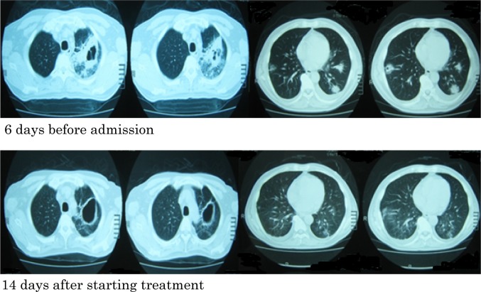
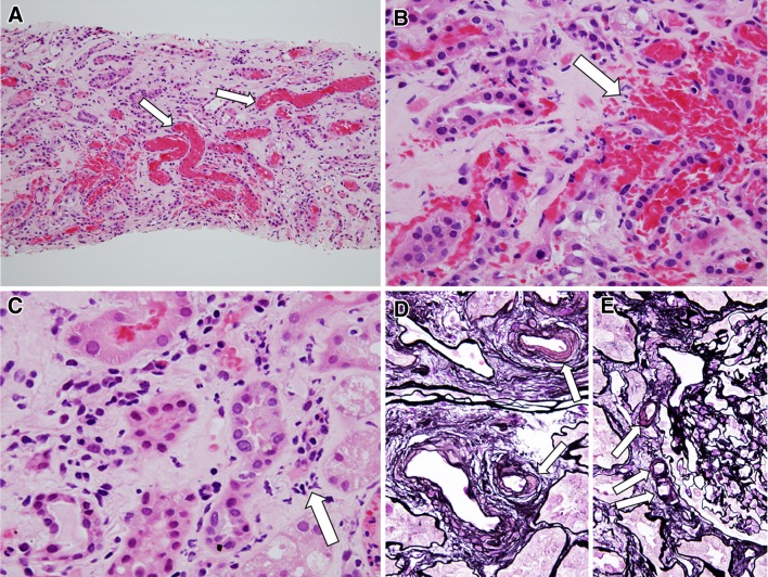
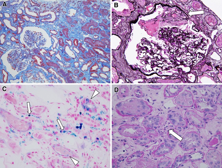
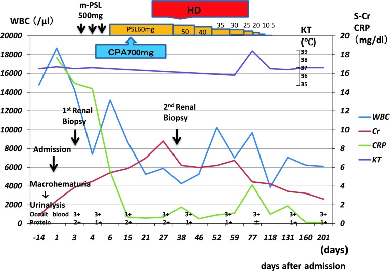
A 60-year-old man had experienced cough, bloody sputum, and a 38 °C fever for 1.5 months. He visited an outpatient clinic and received antibiotics and nonsteroidal anti-inflammatory drugs. However, because the symptoms continued, he visited our hospital. The past medical history included chronic sinusitis, hypertension, and diabetes mellitus. A chest x-ray film and computed tomography showed multiple pulmonary nodules with cavities. Macrohematuria had developed 3 days before admission, and renal function had deteriorated (creatinine, 2.45 mg/dL) in 2 weeks. He was admitted to our hospital because of rapidly progressive glomerulonephritis (RPGN) and multiple pulmonary nodules. On admission, the clinical diagnosis was suspected to be granulomatosis with polyangiitis (Wegener's) (GPA), although tests for proteinase-3 anti-neutrophil cytoplasmic antibody (PR3-ANCA) were negative. Antibiotics were administered for 5 days. After renal biopsy, methylprednisolone pulse therapy and cyclophosphamide pulse therapy were performed. The pathological diagnosis on the basis of the renal biopsy was glomerular and interstitial hemorrhage, possibly associated with vasculitis. After the treatment, the pulmonary symptoms, multiple pulmonary nodules, and severe inflammatory reactions in the peripheral blood were resolved. However, renal dysfunction progressed to end-stage renal disease 1 month after renal biopsy. Hemodialysis was started, and the steroid therapy was continued. During hemodialysis, a second renal biopsy was performed and led to a diagnosis of pauci-immune focal segmental crescentic glomerulonephritis. Renal function gradually recovered, and hemodialysis was discontinued. This case was (double) ANCA-negative GPA which presented prominent glomerular and interstitial hemorrhage, may be associated with small vessel vasculitis, but without active necrotizing and crescentic glomerular lesions, in the rapidly progressive glomerulonephritis.

Keywords: Granulomatosis with polyangiitis (Wegener’s); Proteinase-3 anti-neutrophil cytoplasmic antibody; Rapidly progressive glomerulonephritis.


Figures

Fig. 1
Fig. 1
Upper panel a CT scan obtained 6 days before admission shows consolidation with a cavity in the left upper lobe and multiple areas of consolidation without cavities in both lower lobes. Lower panel a CT scan obtained 14 days after the start of treatment shows that these lesions, other than the cavities, had improved with administration of prednisolone
Fig. 2
Fig. 2
Light microscopic findings of glomeruli from 1st biopsy samples (a, b PAM stain; c, d Masson stain; a ×200; bd: ×600). The biopsy specimens contained 9 glomeruli in which 2 glomeruli were obsolescent. Massive red blood cell casts were present in the renal tubules (arrow in a). In 7 glomeruli, 5 glomeruli showed mild segmental endocapillary proliferation (arrow in b) with infiltration of neutrophils and monocytes. 3 glomeruli showed hemorrhage (arrow in c and d) in Bowman’s space. No necrotizing or crescentic lesions were detected in glomeruli. Immunoflourescence study showed no obvious deposition of immunoglobulin (Ig) G, IgA, IgM, and complement (C)3, C1q, and C4 in glomeruli, indicating pauci-immune type of deposition
Fig. 3
Fig. 3
Light microscopic findings of tubulointerstitium and arterioles from 1st biopsy samples (ac H&E stain; d, e: PAM stain; a ×200; b, c, e ×600; d ×400). In interstitium, many red blood cell casts were present in tubular lumens (arrow in a), may be associated with glomerular hemorrhage. Localized interstitial hemorrhage was also noted with peritubular capillaritis (arrow in b). Peritubular capillaritis was expanded in the renal cortex and medulla with or without interstitial hemorrhage (arrow in c). This patient had DM, and severe hyalinosis of small arterioles (arrow in d), and polar vasculosis around glomerular hilus (arrow in e) were evident although nodular lesions of DM nephropathy could not be detected in glomeruli. No necrotizing or granulomatous vasculitis was noted in arteries
Fig. 4
Fig. 4
Light microscopic findings from 2nd biopsy samples (a Masson stain; b PAM stain; c Berlin blue stain; d H&E stain; a ×200; bd ×600). The biopsy specimens contained 19 glomeruli in which 1 glomerulus showed obsolescence. Interstitial hemorrhage and red blood cell (RBC) casts were disappeared from the renal tubules and interstitium. In the glomeruli, endocapillary proliferative or necrotizing lesions could not be detected, but 3 glomeruli were accompanied by fibrocellular crescents (arrow in b), and 1 glomerulus had fibrous crescent. Berlin blue stain indicated the deposition of hemosiderin in renal tubular epithelial cells (arrowhead in c) and interstitium (arrow in c), indicating that post-status of massive RBC casts in renal tubules and interstitial hemorrhage, respectively. Capillaritis was present in peritubular capillaries (arrow in d), but the degree of capillaritis was milder than 1st biopsy
Fig. 5
Fig. 5
Clinical course of the present case. During the course, macrohematuria was observed only once 3 days before admission, but oliguria was not observed. After 1st renal biopsy, methylprednisolone pulse therapy (500 mg/day for 3 days) and cyclophosphamide pulse therapy (700 mg for 1 day) were administered, but renal dysfunction progressed. So hemodialysis was started, and steroid therapy was continued (60 mg/day for 4 weeks and then tapered by 5–10 mg/week). Renal function gradually recovered, and hemodialysis was discontinued. The most recent measures of renal function showed serum creatinine 2.61 mg/dL, without hemodialysis

References

    1. Guillevin L, Durand-Gasselin B, Cevallos R, Gayraud M, Lhote F, Callard P, et al. Microscopic polyangiitis: clinical and laboratory findings in eighty-five patients. Arthritis Rheum. 1999;42(3):421–430. doi: 10.1002/1529-0131(199904)42:3<421::AID-ANR5>3.0.CO;2-6. - DOI - PubMed
    1. Finkielman JD, Lee AS, Hummel AM, Viss MA, Jacob GL, Homburger HA, et al. ANCA are detectable in nearly all patients with active severe Wegener’s granulomatosis. Am J Med. 2007;120(7):643–14. - PubMed
    1. Falk RJ, Jennette JC. ANCA small-vessel vasculitis. J Am Soc Nephrol. 1997;8(2):314–322. - PubMed
    1. Hagen EC, Daha MR, Hermans J, Andrassy K, Csernok E, Gaskin G, et al. Diagnostic value of standardized assays for anti-neutrophil cytoplasmic antibodies in idiopathic systemic vasculitis. EC/BCR Project for ANCA Assay Standardization. Kidney Int. 1998;53(3):743–753. doi: 10.1046/j.1523-1755.1998.00807.x. - DOI - PubMed
    1. Savige J, Pollock W, Trevisin M. What do antineutrophil cytoplasmic antibodies (ANCA) tell us? Best Pract Res Clin Rheumatol. 2005;19(2):263–276. doi: 10.1016/j.berh.2004.10.003. - DOI - PubMed

LinkOut - more resources