

A case report of the anti-glomerular basement membrane glomerulonephritis with mesangial IgA deposition
- PMID: 28509211
- PMCID: PMC5413720
- DOI: 10.1007/s13730-012-0029-y
A case report of the anti-glomerular basement membrane glomerulonephritis with mesangial IgA deposition
Abstract
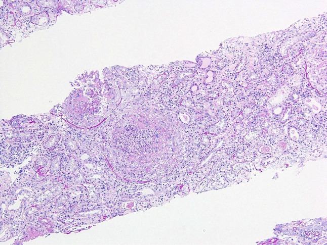
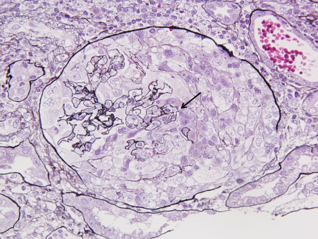
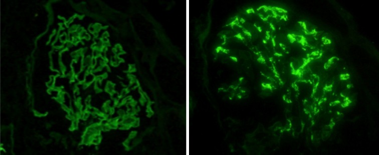
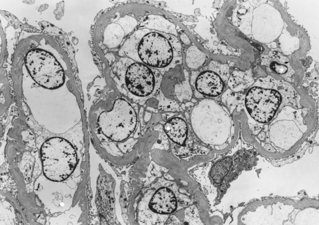
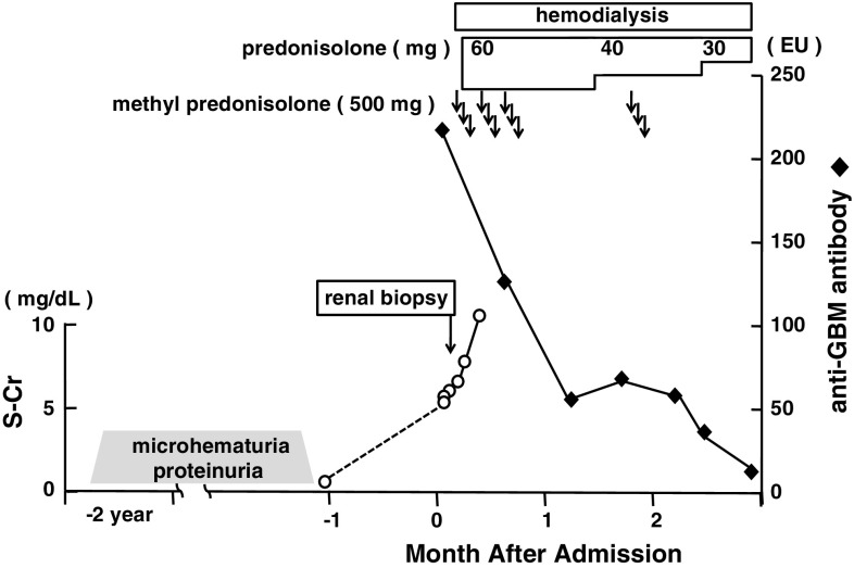
A 46-year-old Japanese male with a past medical history of microscopic hematuria presented with nausea, vomiting, and abdominal pain for which he had been diagnosed with rapidly progressive glomerulonephritis with a peak serum creatinine of 6.6 mg/dL and anti-glomerular basement membrane antibody of 214 EU. Light microscopy showed cellular crescent formation, and immunofluorescence illustrated both linear staining of IgG along the glomerular basement membrane and granular staining of IgA and C3 in the mesangial area; however, the PAS staining of mesangial expansions and mesangial proliferations were not observed. Clinical and histological findings suggested anti-glomerular basement membrane glomerulonephritis with mesangial IgA deposition, suggesting IgA nephropathy, a rare condition.
Keywords: Anti-glomerular basement membrane glomerulonephritis; IgA nephropathy; Rapidly progressive glomerulonephritis.
Figures
References
-
- Jennette JC, Nickeleit V. Anti-glomerular basement membrane glomerulonephritis and Goodpasture’s syndrome. In: Jenette JC, Olson JL, Schwartz MM, Silva FG, editors. Heptinstall’s pathology of the kidney. 6. Philadelphia: Lippincott Williams and Wilkins; 2007. pp. 626–629.
-
- Haas M. IgA nephropathy and Henoch–Schönlein purpura nephritis. In: Jennette JC, Olson JL, Schwartz MM, Silva FG, editors. Heptinstall’s pathology of the kidney. 6. Philadelphia: Lippincott-Williams and Wilkins; 2007. pp. 424–486.
LinkOut - more resources
Full Text Sources
Miscellaneous
