. 2013 Nov;2(2):197-203.
doi: 10.1007/s13730-013-0064-3. Epub 2013 Feb 26.

Proliferative glomerulonephritis with monoclonal IgG2κ deposit successfully treated with steroids: a case report and review of the literature

Affiliations

Proliferative glomerulonephritis with monoclonal IgG2κ deposit successfully treated with steroids: a case report and review of the literature

Ryuji Ohashi et al. CEN Case Rep. 2013 Nov.

Abstract

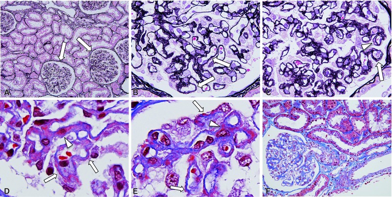
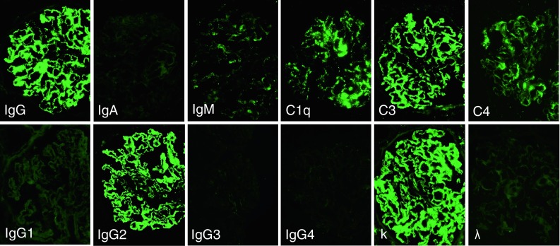
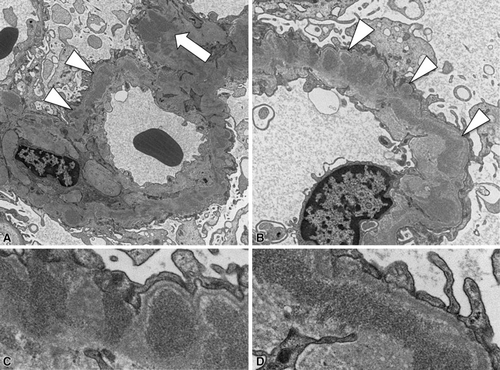
A novel form of glomerular injury with monoclonal immunoglobulin (Ig) IgG deposition, termed "proliferative glomerulonephritis (GN) with monoclonal IgG deposits" (PGNMID), is a recently described entity. PGNMID presents with various histological patterns, such as membranoproliferative GN, endocapillary proliferative GN and membranous nephropathy (MN). The deposits are composed of monoclonal immunoglobulin, most commonly IgG3 and occasionally IgG2. At present, the clinical significance of each IgG subclass and the morphological patterns of glomerular injury have not been fully investigated due to the limited number of PGNMID cases reported. The patient was a 27-year-old woman presenting with a mild degree of proteinuria and no other physical or serological abnormalities. Monoclonal Ig could not be identified in her serum or urine. Renal biopsy found features of MN with deposition of monoclonal IgG2κ. Electron microscopy examination revealed non-organised electron-dense deposits predominantly in subepithelial locations. Based on a diagnosis of PGNMID, she was treated with prednisolone and proteinuria significantly decreased in less than 4 weeks. Although the clinical outcomes of PGNMID remain to be defined, MN features may possibly be a sign of favourable prognosis-a hypothesis supported by recent reports. The absence of advanced chronic damage in the kidney, such as glomerulosclerosis or tubulointerstitial fibrosis, may also have contributed to the favourable outcome in the present case. Further studies on additional PGNMID cases that allow the correlation of morphological features and IgG subclasses with clinical outcomes are needed in order to confirm our findings and further solidify the clinical aspects of this new disease entity.

Keywords: Membranous nephropathy; Monoclonal IgG; Proliferative glomerulonephritis; Steroid.


Figures

Fig. 1
Fig. 1
Light microscopy findings (ac PAM stain, df Masson trichrome stain; a ×200, b, c ×800, d, e ×1600, f ×400). The glomeruli were slightly enlarged (arrows in a), with segmental to global mild thickening of the peripheral capillary walls. Note the segmental to diffuse distribution of irregular spike formations (arrows in b and c), with variably sized subepithelial deposits (arrows in d and e). Double contour of the glomerular basement membranes with intramembranous deposits was noted focally (arrowhead in c). Mesangial deposits were also present in some mesangial areas (arrowheads in d and e). Tubulointerstitial and arterial alterations were not evident (f)
Fig. 2
Fig. 2
Immunofluorescence findings. Immunofluorescence analysis was performed using fluorescein isothiocyanate (FITC)-conjugated antibodies against IgG, IgA, IgM, C3, C1q and C4 (MBL, Nagoya, Japan), and IgG1, IgG2, IgG3 and IgG4 (Binding Site Limited, Birmingham, UK), as well as kappa (κ) and lambda (λ) light chains (Dako). Fine granular staining of IgG (3+) and C3 (3+) was identified, mainly along the glomerular capillary walls and segmentally in the mesangial areas. Of all the IgG subclasses, only IgG2 was significantly positive (3+), while the other IgG subclasses (IgG1, IgG3, IgG4) were all negative. κ light chain was positive (3+) in a manner similar to IgG2, whereas λ light chain was negative. These findings indicate the presence of IgG2κ monoclonal immunoglobulin deposition in the glomeruli. The deposition of IgM (1+), C1q (1+) and C4 (1+) was also noted in segmental peripheral and mesangial areas
Fig. 3
Fig. 3
Electron microscopy findings (a ×7000, b ×12000, c, d ×40000). Electron microscopy revealed irregularly sized EDD predominantly in the intramembranous (arrowheads in a) and subepithelial regions covered by newly formed membranous material (arrowheads in b), and occasionally in mesangial (arrow in a) locations. c, d are high-magnification images of b. No apparent organised structures were noted within these deposits (c, d)

Similar articles

Cited by

References

    1. Ronco P, Plaisier E, Mougenot B, Aucouturier P. Immunoglobulin light (heavy)-chain deposition disease: from molecular medicine to pathophysiology-driven therapy. Clin J Am Soc Nephrol. 2006;1:1342–1350. doi: 10.2215/CJN.01730506. - DOI - PubMed
    1. Joh K. Pathology of glomerular deposition diseases. Pathol Int. 2007;57:551–565. doi: 10.1111/j.1440-1827.2007.02139.x. - DOI - PubMed
    1. Kiremitci S, Calayoglu R, Ensari A, Erbay B. Pathologist’s puzzle: membranoproliferative glomerulonephritis-like features in cryoglobulinemic glomerulonephritis. Pathol Res Pract. 2012;208:254–258. doi: 10.1016/j.prp.2012.01.006. - DOI - PubMed
    1. D’Amico G, Fornasieri A. Cryoglobulinemic glomerulonephritis: a membranoproliferative glomerulonephritis induced by hepatitis C virus. Am J Kidney Dis. 1995;25:361–369. doi: 10.1016/0272-6386(95)90095-0. - DOI - PubMed
    1. Korbet SM, Schwartz MM, Lewis EJ. Immuotactoid glomerulopathy (fibrillary glomerulonephritis) Clin J Am Soc Nephrol. 2006;1:1351–1356. doi: 10.2215/CJN.01140406. - DOI - PubMed

LinkOut - more resources