

Metabolic Portraits of Breast Cancer by HR MAS MR Spectroscopy of Intact Tissue Samples
- PMID: 28509845
- PMCID: PMC5487989
- DOI: 10.3390/metabo7020018
Metabolic Portraits of Breast Cancer by HR MAS MR Spectroscopy of Intact Tissue Samples
Abstract
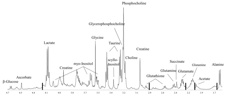
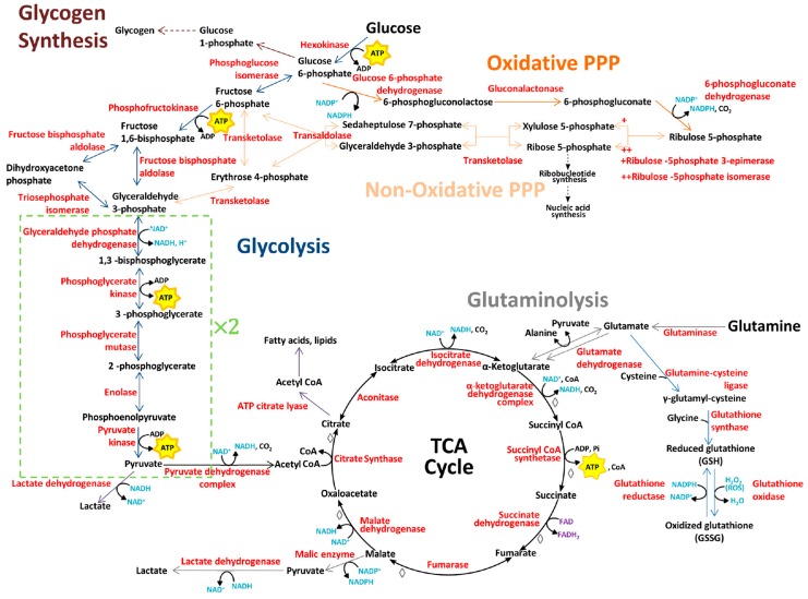
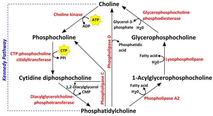
Despite progress in early detection and therapeutic strategies, breast cancer remains the second leading cause of cancer-related death among women globally. Due to the heterogeneity and complexity of tumor biology, breast cancer patients with similar diagnosis might have different prognosis and response to treatment. Thus, deeper understanding of individual tumor properties is necessary. Cancer cells must be able to convert nutrients to biomass while maintaining energy production, which requires reprogramming of central metabolic processes in the cells. This phenomenon is increasingly recognized as a potential target for treatment, but also as a source for biomarkers that can be used for prognosis, risk stratification and therapy monitoring. Magnetic resonance (MR) metabolomics is a widely used approach in translational research, aiming to identify clinically relevant metabolic biomarkers or generate novel understanding of the molecular biology in tumors. Ex vivo proton high-resolution magic angle spinning (HR MAS) MR spectroscopy is widely used to study central metabolic processes in a non-destructive manner. Here we review the current status for HR MAS MR spectroscopy findings in breast cancer in relation to glucose, amino acid and choline metabolism.
Keywords: HR MAS MR spectroscopy; amino acid metabolism; breast cancer; choline phospholipids; glycolysis.
Conflict of interest statement
The authors declare no conflict of interest.
Figures
References
-
- Cancer Registry of Norway . Cancer In Norway 2015—Cancer Incidence, Mortality, Survival and Prevalence In Norway. Cancer Registry of Norway; Oslo, Norway: 2016.
-
- Sørlie T., Perou C.M., Tibshirani R., Aas T., Geisler S., Johnsen H., Hastie T., Eisen M.B., van de Rijn M., Jeffrey S.S., et al. Gene expression patterns of breast carcinomas distinguish tumor subclasses with clinical implications. Proc. Natl. Acad. Sci. USA. 2001;98:10869–10874. doi: 10.1073/pnas.191367098. - DOI - PMC - PubMed
Publication types
LinkOut - more resources
Full Text Sources
Other Literature Sources
Molecular Biology Databases
