Review
. 2010 Feb;2(1):29-38.
doi: 10.1007/s12551-009-0027-2. Epub 2009 Dec 18.

New factors contributing to dynamic calcium regulation in the skeletal muscle triad-a crowded place

Affiliations
Review

New factors contributing to dynamic calcium regulation in the skeletal muscle triad-a crowded place

Oliver Friedrich et al. Biophys Rev. 2010 Feb.

Abstract

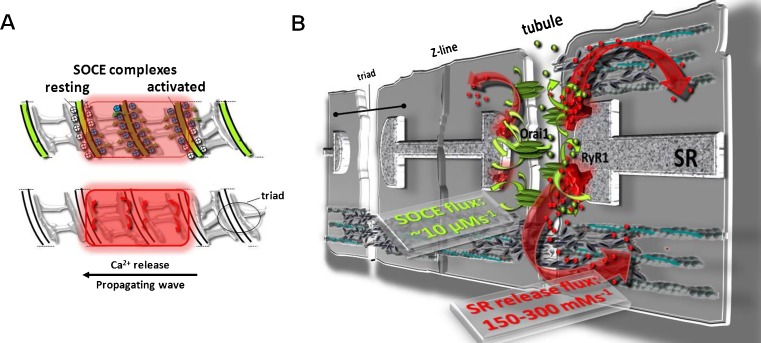
Skeletal muscle is a highly organized tissue that has to be optimized for fast signalling events conveying electrical excitation to contractile response. The site of electro-chemico-mechanical coupling is the skeletal muscle triad where two membrane systems, the extracellular t-tubules and the intracellular sarcoplasmic reticulum, come into very close contact. Structure fits function here and the signalling proteins DHPR and RyR1 were the first to be discovered to bridge this gap in a conformational coupling arrangement. Since then, however, new proteins and more signalling cascades have been identified just in the last decade, adding more diversity and fine tuning to the regulation of excitation-contraction coupling (ECC) and control over Ca2+ store content. The concept of Ca2+ entry into working skeletal muscle has become attractive again with the experimental evidence summarized in this review. Store-operated Ca2+ entry (SOCE), excitation-coupled Ca2+ entry (ECCE), action-potential-activated Ca2+ current (APACC), and retrograde EC-coupling (ECC) are new concepts additional to the conventional orthograde ECC; they have provided fascinating new insights into muscle physiology. In this review, we discuss the discovery of these pathways, their potential roles, and the signalling proteins involved that show that the triad may become a crowded place in time.

Keywords: Action-potential-activated Ca2+ entry; Ca2+ sparks; DHPR; Excitation-coupled Ca2+ entry; Retrograde coupling; RyR; Skeletal muscle; Store-operated Ca2+ entry; T-system; Triad.


Figures

Fig. 1a, b
Fig. 1a, b
Conformational coupling of SOCE complexes in adult skeletal muscle. a Model of locally controlled EC-coupling during a low Mg2+-induced Ca2+ wave propagating along the fiber axis in a mechanically skinned fiber with Fluo-5N trapped in the t-system and rhod-2 in the cytoplasm. SOCE can be detected with a “coupling delay” of ∼25 ms from local SR release (according to data from Launikonis et al. 2009a, b). b Proposed model of Orai1 channels bridging the “Ca2+ supply gap” that may be caused by Ca2+ during the global release not being immediately available for SR refilling due to buffering by cytoplasmic buffers. SOCE may provide a shortcut for Ca2+ being directly transferred from the tubules through the triad junction into the SR and maintains SR filling during sustained physiological activity

References

    1. Allen DG, Lamb GD, Westerblad H. Skeletal muscle fatigue: cellular mechanisms. Physiol Rev. 2008;88:287–332. doi: 10.1152/physrev.00015.2007. - DOI - PubMed
    1. Allard B, Couchoux H, Pouvreau S, Jacquemond V. Sarcoplasmic reticulum Ca2+ release and depletion fail to affect sarcolemmal ion channel activity in mouse skeletal muscle. J Physiol. 2006;575(1):68–81. doi: 10.1113/jphysiol.2006.112367. - DOI - PMC - PubMed
    1. Andronache Z, Hamilton SL, Dirksen RT, Melzer W. A retrograde signal from RyR1 alters DHP receptor inactivation and limits window Ca2+ release in muscle fibers of Y522S RyR1 knock-in mice. Proc Natl Acad Sci USA. 2009;106:4531–4536. doi: 10.1073/pnas.0812661106. - DOI - PMC - PubMed
    1. Bannister RA. Bridging the myoplasmic gap: recent developments in skeletal muscle excitation-contraction coupling. J Muscle Res Cell Motil. 2007;28:275–283. doi: 10.1007/s10974-007-9118-5. - DOI - PubMed
    1. Bannister RA, Pessah IN, Beam KG. The skeletal L-type Ca2+ current is a major contributor to excitation-coupled Ca2+ entry. J Gen Physiol. 2009;133(1):79–91. doi: 10.1085/jgp.200810105. - DOI - PMC - PubMed

LinkOut - more resources