. 2015 Mar;7(1):5-14.
doi: 10.1007/s12551-014-0151-5. Epub 2014 Dec 20.

The role of super-relaxed myosin in skeletal and cardiac muscle

Affiliations

The role of super-relaxed myosin in skeletal and cardiac muscle

James W McNamara et al. Biophys Rev. 2015 Mar.

Abstract

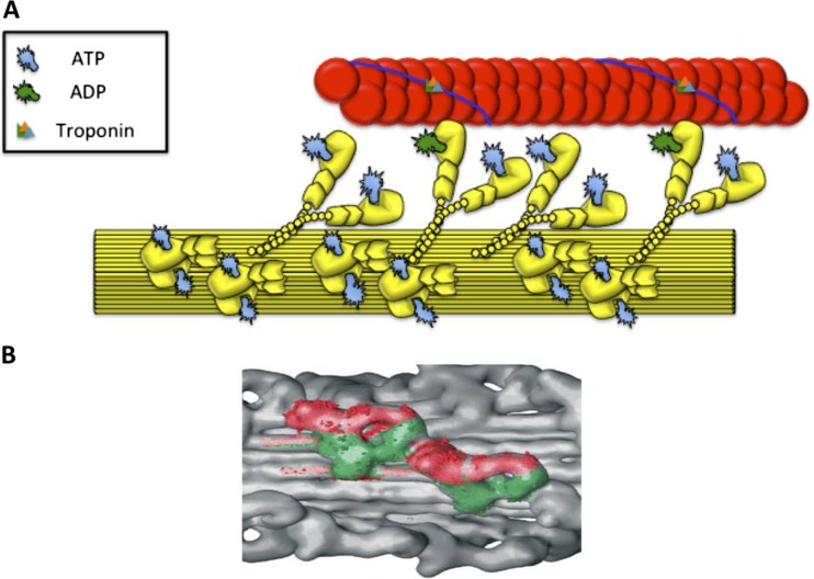
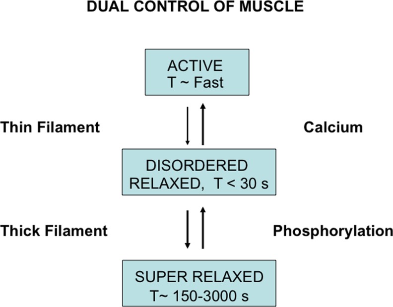
The super-relaxed (SRX) state of myosin was only recently reported in striated muscle. It is characterised by a sub-population of myosin heads with a highly inhibited rate of ATP turnover. Myosin heads in the SRX state are bound to each other along the thick filament core producing a highly ordered arrangement. Upon activation, these heads project into the interfilament space where they can bind to the actin filaments. Thus far, the population and lifetimes of myosin heads in the SRX state have been characterised in rabbit cardiac, and fast and slow skeletal muscle, as well as in the skeletal muscle of the tarantula. These studies suggest that the role of SRX in cardiac and skeletal muscle regulation is tailored to their specific functions. In skeletal muscle, the SRX modulates the resting metabolic rate. Cardiac SRX represents a "reserve" of inactive myosin heads that may protect the heart during times of stress, e.g. hypoxia and ischaemia. These heads may also be called up when there is a sustained demand for increased power. The SRX in cardiac muscle provides a potential target for novel therapies.

Keywords: ATP; Heart disease; Myosin heads; Super-relaxation.


Figures

Fig. 1
Fig. 1
a The three states of myosin in striated muscle: active, disordered relaxed and super-relaxed. Active myosin produces force by pulling the actin filaments (red) towards the M-line of the sarcomere. Relaxed myosin in the disordered state protrudes into the interfilament space but is restricted from binding to actin by the thin filament regulatory proteins whilst myosin in the super-relaxed (SRX) state is bound on or close to the thick filament. This results in an ATP turnover lifetime that is at least 5-fold longer in SRX myosin than disordered myosin. b An atomic model of relaxed myosin heads bound to the thick filament core. In the SRX state the blocked (green) and free (red) myosin heads interact with each other forming the J-like structure of the interacting heads motif. These heads may also bind to the thick filament core (grey). (b) reproduced with permission from Craig and Woodhead (2006)
Fig. 2
Fig. 2
The three states of myosin in striated muscle. Active myosin has a rapid ATP turnover lifetime of under 1 s, while disordered relaxed myosin is generally less than 30 s. Activation of these myosin heads may occur through calcium binding to the thin filament regulatory proteins. The super-relaxed myosin has a highly inhibited ATP turnover time i.e. longer than 100 s. Phosphorylation of the thick filament proteins cMyBP-C and RLC may facilitate the transfer of myosin heads out of this state. Modified from Cooke (2011)
Fig. 3
Fig. 3
The change in fluorescence intensity as a function of time. The red trace shows the decay seen when 250 μM MANT-ATP is chased with 4 mM ATP. The blue trace shows the inverse experiment where the fibre was incubated with 4 mM ATP and chased with 250 μM MANT-ATP. The change in fluorescence intensity has two phases, a fast phase over the first 30 s and a slow phase over the next ~600 s. This second phase is credited to the slow release of nucleotides from the fraction of myosin heads in the super-relaxed state. Reproduced with permission from Cooke (2011)
Fig. 4
Fig. 4
Changes in the SRX state of cardiac and skeletal muscle fibres in ADP and Ca2+ chases. a The effect of ADP on the skeletal muscle SRX. The open circles show MANT-ATP chased by ATP, while the open triangles indicate MANT-ATP chased by ADP, where the slow decay of fluorescence intensity eliminated. The squares indicate the change in fluorescence intensity from an ATP incubation chased by MANT-ATP. b Effect of ADP on cardiac muscle SRX. Filled circles show the decay of fluorescence intensity in an ATP chase, whereas the open circles show the decay in response to ADP chase, where the SRX is diminished but still present. The open triangles show the change in fluorescence intensity when MANT-ADP is chased with ADP. c, d The effect of calcium activation on skeletal (c) and cardiac (d) muscle fibres. Red shows the decay in fluorescence intensity in ATP chase experiments, while the blue traces are the calcium activation chase (ATP plus calcium, pCa = 5.7). Skeletal SRX was lost upon activation while cardiac SRX was largely unchanged. (a) reproduced with permission from Stewart et al. (2010). (bd) reproduced with permission from Hooijman et al. (2011)

References

    1. Alamo L, Wriggers W, Pinto A, Bártoli F, Salazar L, Zhao F-Q, Craig R, Padrón R. Three-dimensional reconstruction of tarantula myosin filaments suggests how phosphorylation may regulate myosin activity. J Mol Biol. 2008;384:780–797. doi: 10.1016/j.jmb.2008.10.013. - DOI - PMC - PubMed
    1. AL-Khayat HA, Kensler RW, Squire JM, Marston SB, Morris EP. Atomic model of the human cardiac muscle myosin filament. Proc Natl Acad Sci U S A. 2013;110:318–323. doi: 10.1073/pnas.1212708110. - DOI - PMC - PubMed
    1. Allen DG, Kentish JC. The cellular basis of the length-tension relation in cardiac muscle. J Mol Cell Cardiol. 1985;17:821–840. doi: 10.1016/S0022-2828(85)80097-3. - DOI - PubMed
    1. Ashrafian H, Redwood C, Blair E, Watkins H. Hypertrophic cardiomyopathy:a paradigm for myocardial energy depletion. Trends Genet. 2003;19:263–268. doi: 10.1016/S0168-9525(03)00081-7. - DOI - PubMed
    1. Barefield D, Sadayappan S. Phosphorylation and function of cardiac myosin binding protein-C in health and disease. J Mol Cell Cardiol. 2010;48:866–875. doi: 10.1016/j.yjmcc.2009.11.014. - DOI - PMC - PubMed

LinkOut - more resources