. 2015 Mar;7(1):63-76.
doi: 10.1007/s12551-014-0157-z. Epub 2015 Jan 21.

Phospholamban phosphorylation, mutation, and structural dynamics: a biophysical approach to understanding and treating cardiomyopathy

Affiliations

Phospholamban phosphorylation, mutation, and structural dynamics: a biophysical approach to understanding and treating cardiomyopathy

Naa-Adjeley D Ablorh et al. Biophys Rev. 2015 Mar.

Abstract

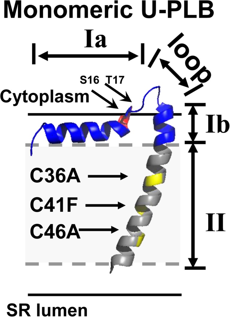
We review the recent development of novel biochemical and spectroscopic methods to determine the site-specific phosphorylation, expression, mutation, and structural dynamics of phospholamban (PLB), in relation to its function (inhibition of the cardiac calcium pump, SERCA2a), with specific focus on cardiac physiology, pathology, and therapy. In the cardiomyocyte, SERCA2a actively transports Ca2+ into the sarcoplasmic reticulum (SR) during relaxation (diastole) to create the concentration gradient that drives the passive efflux of Ca2+ required for cardiac contraction (systole). Unphosphorylated PLB (U-PLB) inhibits SERCA2a, but phosphorylation at S16 and/or T17 (producing P-PLB) changes the structure of PLB to relieve SERCA2a inhibition. Because insufficient SERCA2a activity is a hallmark of heart failure, SERCA2a activation, by gene therapy (Andino et al. 2008; Fish et al. 2013; Hoshijima et al. 2002; Jessup et al. 2011) or drug therapy (Ferrandi et al. 2013; Huang 2013; Khan et al. 2009; Rocchetti et al. 2008; Zhang et al. 2012), is a widely sought goal for treatment of heart failure. This review describes rational approaches to this goal. Novel biophysical assays, using site-directed labeling and high-resolution spectroscopy, have been developed to resolve the structural states of SERCA2a-PLB complexes in vitro and in living cells. Novel biochemical assays, using synthetic standards and multidimensional immunofluorescence, have been developed to quantitate PLB expression and phosphorylation states in cells and human tissues. The biochemical and biophysical properties of U-PLB, P-PLB, and mutant PLB will ultimately resolve the mechanisms of loss of inhibition and gain of inhibition to guide therapeutic development. These assays will be powerful tools for investigating human tissue samples from the Sydney Heart Bank, for the purpose of analyzing and diagnosing specific disorders.

Keywords: Loss-of-inhibition mutants; Phospholamban; Phosphorylation; SERCA2a; Subunit model.


Figures

Fig. 1
Fig. 1
Therapeutic strategies based on two models of SERCA2a/PLB regulation. Fluorescence resonance energy transfer (yellow arrows) from a donor fluorophore (yellow sun, indicating emission) bound to SERCA2a (blue) to an acceptor fluorophore (orange triangle) bound to PLB (red) is indicated by decreased emission (smaller yellow sun). The circled P is phosphate and the purple diamond is a drug. Red PLB is endogenous PLB (WT-PLB) and green PLB is mutant PLB. a Dissociation model: PLB phosphorylation causes PLB to dissociate from SERCA2a, thus relieving inhibition, which should eliminate FRET. Therapeutic strategies: decrease PLB expression (not shown), displace PLB from SERCA2a with drug, or increase PLB phosphorylation. b Subunit model: PLB is essentially a subunit of SERCA2a. Phosphorylation causes PLB to adopt a more extended structure (R-state) without dissociation from SERCA2a, which should increase FRET. Therapeutic strategies include stabilizing PLB R-state with drug, or using gene therapy to deliver a loss-of-inhibition PLB mutant that binds to SERCA2a, thus displacing WT PLB
Fig. 2
Fig. 2
PLB antibody affinities. Top Western blots of purified synthetic U-PLB and P16-PLB (5, 10, and 15 ng) on the same membrane, blotted with one of three PLB antibodies: Ab8a3, Ab2D12, or AbA1. The intensities were measured using a box that encompassed all of the oligomeric states of PLB (magenta box on blot Ab8a3). Bottom Intensities of the 5, 10 and 15 ng of U-PLB (circles) and P16-PLB (squares) versus their concentration and the resulting standard curves from the blot labeled with the same PLB antibody (εu = the slope of the standard curve for U-PLB εp = the slope of the standard curve for P-PLB). This figure was adapted from Ablorh et al. (2012) with permission from Elsevier
Fig. 3
Fig. 3
The intensities of mixtures of synthetic U-PLB and synthetic P16-PLB with P16-PLB mole fractions of 0.0, 0.25, 0.50, 0.75, and 1.00 obtained from western blots with either antibody Ab8a3 (magenta square), Ab2D12 (blue circle), or AbA1 (green triangle). Values are means ± SEM. All mixtures contain 12 ng of total PLB. If the antibodies bound U-PLB and P16-PLB with equal affinity, the intensities of the mixtures would be equal (independent of the P16-PLB mole fraction). Instead, the intensities of the mixtures decrease as the mole fraction of P16-PLB increases. This indicates that the PLB antibodies bind U-PLB with greater affinity than they bind P16-PLB, and that none of the 3 PLB antibodies can be used to accurately determine PLB expression in a single blot
Fig. 4
Fig. 4
a Validation of the method: four identical western blots containing 2.5, 5, and 11 ng of synthetic U-PLB (U), P16-PLB (P16), P17-PLB (P17), and 2P-PLB (2P), and mixtures of standards (ae). blotted with 4 different PLB antibodies (AbU, Ab17, Ab16, and Ab2P). As in Fig. 2, all oligomeric bands were included in the intensity measurements. b Application of the method: mole fraction of each PLB phosphorylation state in pig tissue homogenates with (solid) and without (open) left ventricular hypertrophy (mean ± SEM). The figure was reproduced from Ablorh et al. (2014) with permission from the American Society of Biological Chemistry and Molecular Biology
Fig. 5
Fig. 5
SERCA2a activity. a The enzyme-linked NADH assay for measuring SERCA2a activity and calculating ∆pKCa. SERCA2a ATPase activity is coupled to phosphorylation of pyruvate by pyruvate kinase. NADH oxidation is coupled to dehydrogenation of pyruvate to form lactate. The oxidation of NADH is measured by absorbance at 280 nm. b ∆pKCa of U-PLB (U), P16-PLB (P16), P17-PLB (P17), and 2P-PLB (2P) with the reference point of pKCa for SERCA2a alone. The figure was reproduced from Ablorh et al. with permission from the American Society of Biological Chemistry and Molecular Biology
Fig. 6
Fig. 6
PLB equilibria. In the T-state/R-state equilibrium, the T-state dominates in both monomeric and pentameric PLB. In the monomer/pentamer equilibria, the pentamer dominates in both the T-state and R-state equilibrium so that pentameric, T-state PLB is the dominant form
Fig. 7
Fig. 7
AFA-U-PLB shows PLB dynamic domains with identification of phosphorylation sites (S16 in red and T17 in dark red) and sites for monomeric mutations (C36A, C41F, and C46A in yellow). Gray dashes outline the hydrophobic core in the phospholipid bilayer. This structure is adapted from frame 12 of PDB file 2 KB7 (Traaseth et al. 2009). Sequence of human PLB: M E K V Q Y L T R S A I R R A S T I E M P Q Q A R Q K L Q N L F I N F C L I L I C L L L I C I I V M L L
Fig. 8
Fig. 8
2-color SERCA2a used for small-molecule screening. a Computational model of GFP and RFP modeled on the crystal structure of SERCA2a1a (PDB 1IWO). b Confocal imaging of live HEK cells expressing 2-color SERCA2a. Yellow co-localization of RFP and GFP fluorescence, Blue DAPI nuclear stain. Thus, 2-color SERCA2a is localized to intracellular ER membranes. This figure is reproduced from Gruber et al. (2014) with permissions from Sage Publishing
Fig. 9
Fig. 9
FRET competition in live cells (Gruber et al. 2012). a FRET is detected from CFP-SERCA2a to YFP-PLB, expressed stably in HEK cells (left), but transient transfection with unlabeled PLB mutant (PLB M, green) displaces YFP-PLB, eliminating FRET and relieving SERCA2a inhibition. b FRET efficiency E (error bars ±SEM). AID (activation-induced deaminase, a cytoplasmic protein) is a transfection control to rule out non-specific transfection artifacts. Decreased FRET (E) between SERCA2a and YFP-PLB in the presence of the unlabeled mutants (PLBM) indicates that unlabeled PLBM constructs compete effectively with YFP-PLB for binding to CFP-SERCA2a. Adapted from Gruber et al. (2012) with permission from Elsevier
Fig. 10
Fig. 10
Two mechanisms to relieve SERCA2a inhibition by PLB in cardiac SR. Phosphorylation shifts the PLB cytoplasmic domain toward the dynamically disordered R state. Micromolar Ca2+ induces a structural change in the SERCA2a transmembrane domain. The figure was reproduced from Dong and Thomas (2014) with permission from Elsevier

References

    1. Ablorh NA, Miller T, Nitu F, Gruber SJ, Karim C, Thomas DD. Accurate quantitation of phospholamban phosphorylation. Immunoblot Anal Biochem. 2012;425:68–75. doi: 10.1016/j.ab.2012.01.028. - DOI - PMC - PubMed
    1. Ablorh NA, Dong X, James ZM, Xiong Q, Zhang J, Thomas DD, Karim CB. Synthetic Phosphopeptides Enable Quantitation of the Content and Function of Phospholamban's four Phosphorylation States. Cardiac Muscle J Biol Chem. 2014 - PMC - PubMed
    1. Abu-Baker S, Lorigan GA. Phospholamban and its phosphorylated form interact differently with lipid bilayers: A 31P, 2H, and 13C solid-state NMR spectroscopic study. Biochemistry. 2006;45:13312–13322. doi: 10.1021/bi0614028. - DOI - PMC - PubMed
    1. Afara MR, Trieber CA, Glaves JP, Young HS. Rational design of peptide inhibitors of the sarcoplasmic reticulum calcium pump. Biochemistry. 2006;45:8617–8627. doi: 10.1021/bi0523761. - DOI - PubMed
    1. Andino LM, Takeda M, Kasahara H, Jakymiw A, Byrne BJ, Lewin AS. AAV-mediated knockdown of phospholamban leads to improved contractility and calcium handling in cardiomyocytes. J Gen Med. 2008;10:132–142. doi: 10.1002/jgm.1131. - DOI - PubMed

LinkOut - more resources