

Review
doi: 10.1007/s12551-009-0011-x.
Epub 2009 Jun 6.
Corneal collagen-its role in maintaining corneal shape and transparency
Affiliations
- PMID: 28509987
- PMCID: PMC5425665
- DOI: 10.1007/s12551-009-0011-x
Item in Clipboard
Review
Corneal collagen-its role in maintaining corneal shape and transparency
Biophys Rev.
2009 Jul.
Abstract
Corneal collagen has a number of properties that allow it to fulfil its role as the main structural component within the tissue. Fibrils are narrow, uniform in diameter and precisely organised. These properties are vital to maintain transparency and to provide the biomechanical prerequisites necessary to sustain shape and provide strength. This review describes the structure and arrangement of corneal collagen from the nanoscopic to the macroscopic level, and how this relates to the maintenance of the form and transparency of the cornea.
Keywords: Collagen; Cornea; Structure; Transparency.
Figures
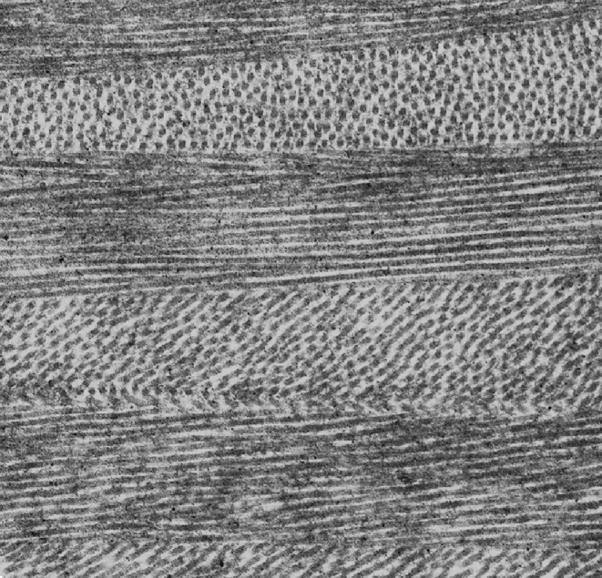
Ultrastructure of the corneal stroma showing the orientation of collagen fibrils within lamellae. Magnification 20K (courtesy of Dr. R. Young)
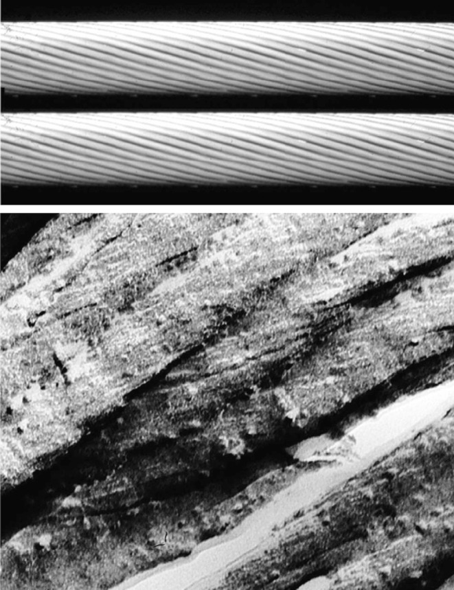
(Above) Molecular model of the sub-fibrillar helicoidal arrangement in corneal collagen. (Below) Freeze-fracture micrograph of hydrated bovine corneal collagen fibrils. Magnification 160K (Ottani et al. , reproduced with permission of the copyright holder)
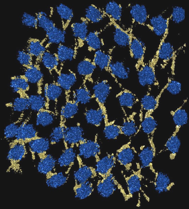
3-D electron microscope reconstruction of the arrangement of proteoglycans stained with Cuprolinic blue dye (yellow) and collagen fibrils in cross-section (blue) in the bovine cornea (courtesy of Dr. C. Knupp and Dr. P. Booth)
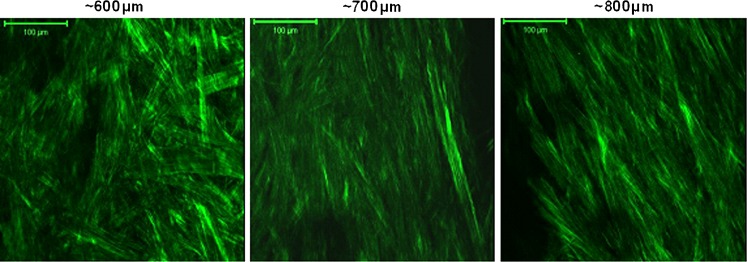
Collagenous lamellae at different depths at the limbus, imaged by second harmonic generated two-photon microscopy. The aligned collagen at greater depths is the limbal annulus (courtesy of Dr. C. Kamma-Lorger)

Wide-angle X-ray scatter patterns taken across the cornea have allowed the preferential arrangement of the corneal lamellae to be mapped. Patterns at specific points around the limbus are shown here superimposed on a schematic of the changes in direction of the lamellae
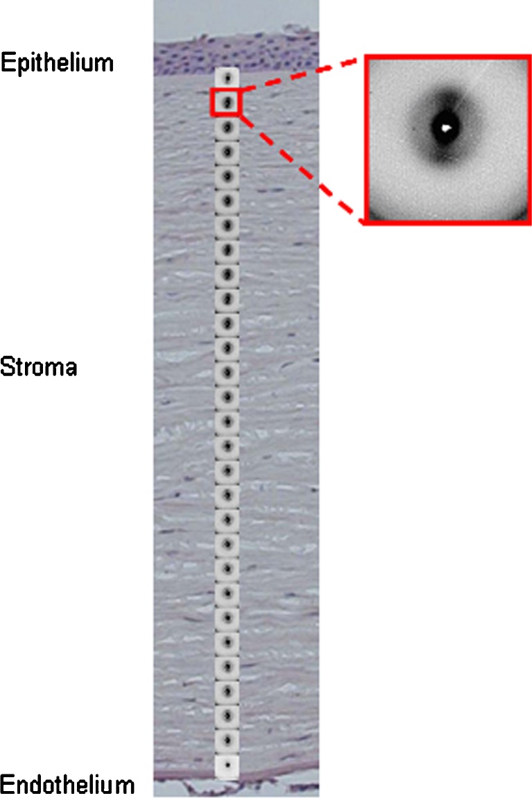
Microfocus X-ray diffraction patterns taken through a thin strip of human cornea from anterior to posterior, shown superimposed on a histological section of a cornea
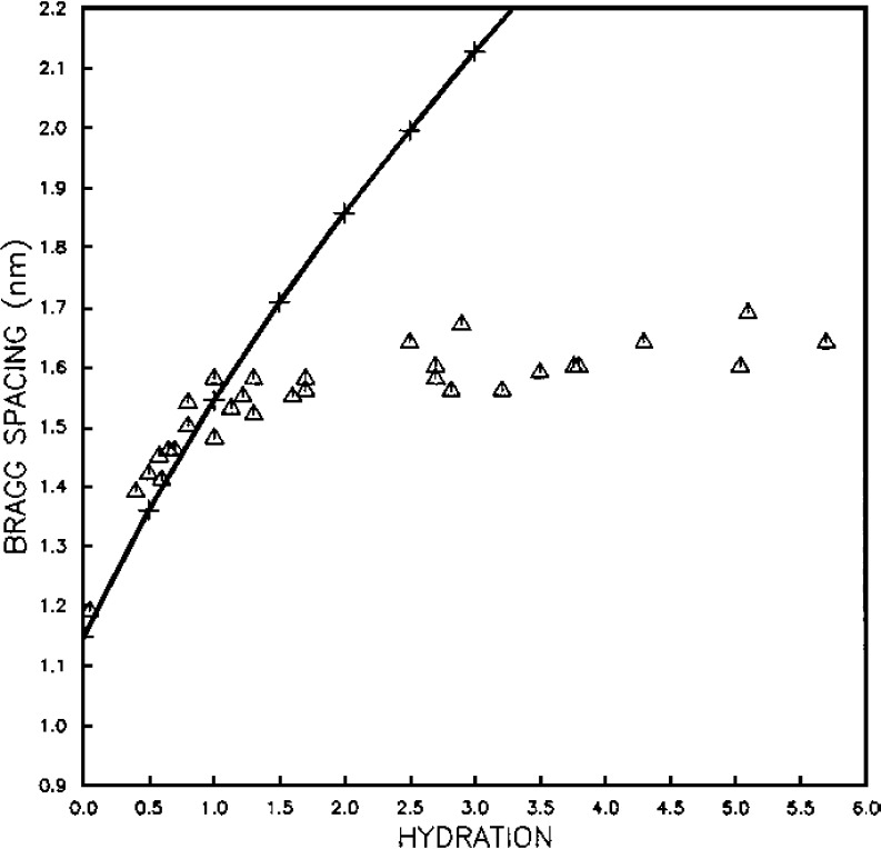
Triangles show the variation of intermolecular Bragg spacing as a function of hydration for bovine cornea. Crosses show the variation of interfibrillar Bragg spacing as a function of hydration, normalised to the intermolecular data at H=0 (reproduced from Meek et al. (1991) with permission of the copyright holder)
References
-
- Aghamohammadzadeh H, Newton RH, Meek KM. X-ray scattering used to map the preferred collagen orientation in the human cornea and limbus. Structure. 2004;12:249–256. - PubMed
-
- Akhtar S, Bron AJ, Salvi SM, Hawksworth NR, Tuft SJ, Meek KM. Ultrastructural analysis of collagen fibrils and proteoglycans in keratoconus. Acta Ophthalmol Scand. 2008;86:764–772. - PubMed
-
- Benedek GB. Theory of transparency of the eye. Appl Opt. 1971;10:459–473. - PubMed
Publication types
Grants and funding
LinkOut - more resources
Full Text Sources
Other Literature Sources
