Review
. 2009 Sep;1(3):149.
doi: 10.1007/s12551-009-0017-4. Epub 2009 Sep 11.

Muscle myosin filaments: cores, crowns and couplings

Affiliations
Review

Muscle myosin filaments: cores, crowns and couplings

John M Squire. Biophys Rev. 2009 Sep.

Abstract

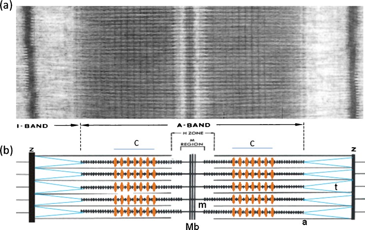
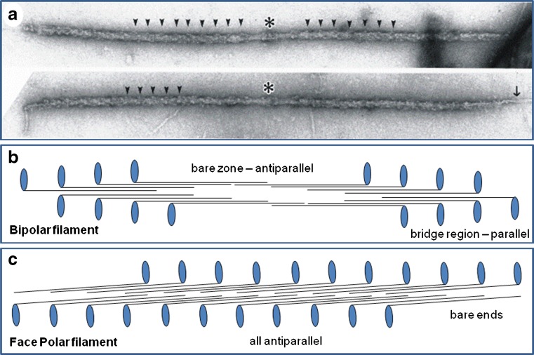
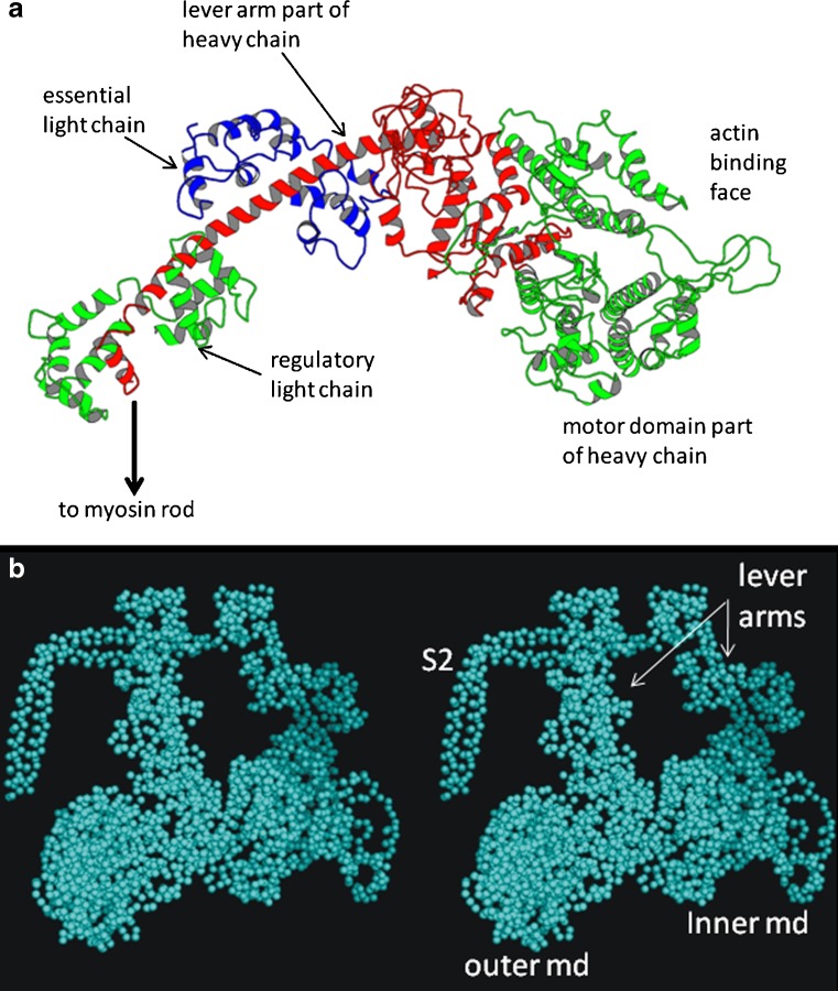
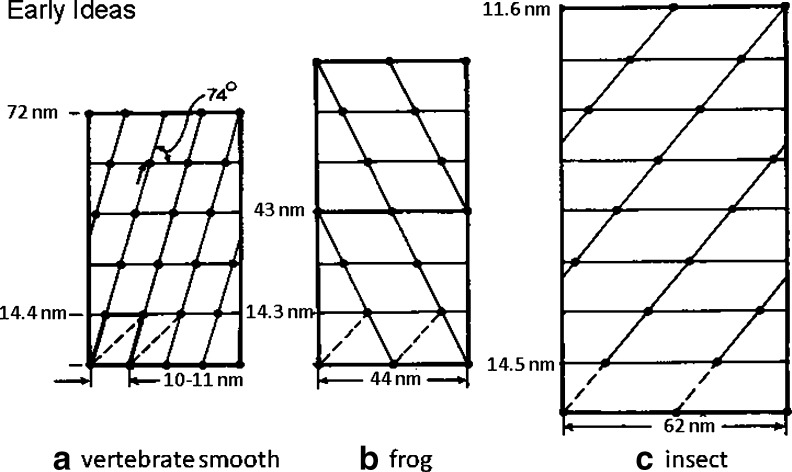
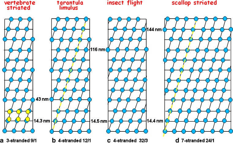
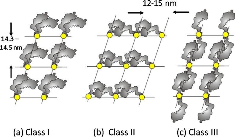
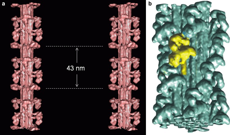
Myosin filaments in muscle, carrying the ATPase myosin heads that interact with actin filaments to produce force and movement, come in multiple varieties depending on species and functional need, but most are based on a common structural theme. The now successful journeys to solve the ultrastructures of many of these myosin filaments, at least at modest resolution, have not been without their false starts and erroneous sidetracks, but the picture now emerging is of both diversity in the rotational symmetries of different filaments and a degree of commonality in the way the myosin heads are organised in resting muscle. Some of the remaining differences may be associated with how the muscle is regulated. Several proteins in cardiac muscle myosin filaments can carry mutations associated with heart disease, so the elucidation of myosin filament structure to understand the effects of these mutations has a clear and topical clinical relevance.

Keywords: Heart disease; Muscle; Myosin filaments; Myosin heads.


Figures

Fig. 1
Fig. 1
a Electron micrograph of a longitudinal section from frog muscle showing the A-band where the bipolar myosin filaments are (see b). The actin filaments run through the I-band from the Z-line and into the A-band where they interdigitate with the myosin filaments. The actin filaments end at the edge if the H-zone. b Schematic diagram of the overlapping myosin (m) and actin (a) filaments. The protein titin (t; blue) runs from the M-band (Mb) along the myosin filaments and then across the I-band to the Z-line (Z). C-protein (MyBP-C) forms a set of stripes in the two halves of the A-band at the positions marked C (orange stripes). C-protein binds to the myosin filament backbone and in some conditions extends out to bind to actin. Figure modified from Fig. 5 of Squire et al. (2005)
Fig. 2
Fig. 2
a Electron micrographs of isolated myosin filaments from bony fish muscle, adapted from AL-Khayat et al. (2008). The M-band is centrally located (asterisk) and some of the 43 nm crown repeats are indicated by arrowheads. The single arrow on the right indicates so-called end filaments which are part of the titin assembly at the tip of the myosin filaments (Trinick 1981). b Schematic diagram showing how myosin molecules pack together to give bipolar myosin filaments. Each myosin molecule is represented as a black line with a blue globular region on one end. Each blue oval represents the myosin head pair of one molecule. Rod packing is antiparallel in the middle of the filaments and parallel at each end. The parallel packing continues for very many crowns on each side of the central bare zone (the head-free region). Myosin molecules in one half of the filament have opposite polarity to those in the other half. This compares with the different packing scheme in (c) for the face polar myosin filaments in vertebrate smooth muscle. Here, the packing is antiparallel throughout and the molecules on opposite faces of the filaments point in opposite directions
Fig. 3
Fig. 3
a Ribbon diagram of the structure of the myosin head determined by Rayment et al. (1993a, b), showing the key components of the head; the motor domain shown in green on the right, the continuation of the heavy chain in red going back to link to the myosin rod, and the two light chains, the essential light chain (blue) and the regulatory light chain (green left). b Stereo pair of a head coupling scheme similar to the Wendt et al. (2001) structure for vertebrate smooth muscle HMM. The motor domain (md) of the inner head (right) binds to the junction between the essential light chain and the back of the motor domain of the outer head. S2 is part of subfragment 2 of the rod part of the myosin molecule. The whole of S2 together with two heads is known as heavy meromyosin (HMM)
Fig. 4
Fig. 4
The early published ideas about the arrangements of myosin heads on myosin filaments in a vertebrate smooth muscle (Small and Squire 1972), b frog sartorius muscle (Huxley and Brown 1967) and c insect (Lethocerus) flight muscle (Reedy 1968). The marked differences in the way that the myosin molecules would have to pack to make these structures prompted Squire (1971) to propose an alternative set of structures for (b) and (c)
Fig. 5
Fig. 5
The accepted symmetries for the myosin filaments in a vertebrate striated muscle, b tarantula and Limulus myosin filaments, c insect flight muscle (Lethocerus) and d scallop striated adductor muscle. These structures all have the myosin head pairs grouped in very similar surface arrays; a common axial repeat of about 14.3–14.5 nm and a common lateral spacing of around 10–15 nm). The slightly different tilt angles of the helical strands give rise to the different observed long repeats. The yellow dashed lines in (b) and (d) indicate the long-pitched helices along which the Class III head interactions (Fig. 6) were originally thought to occur
Fig. 6
Fig. 6
The three Classes of head coupling described by Squire et al. (2005). Invertebrate myosin filaments were originally thought to be Class III, with heads interacting between adjacent crowns along the long-pitched helices shown in the helical nets in Fig. 4. Now, they have been shown to be Class I structures (e.g. Woodhead et al. ; heads within the same myosin molecule interacting). The Class II structure with heads from two different myosin molecules interacting around a single crown has been proposed for insect flight muscle myosin filaments (Lethocerus; AL-Khayat et al. 2003). An unlikely fourth possibility is that the heads do not interact with each other at all (Class IV)
Fig. 7
Fig. 7
Recent images of myosin filament models from a vertebrate striated muscle determined by modelling the low-angle X-ray diffraction pattern from bony fish muscle and shown in stereo (AL-Khayat and Squire 2006) with the backbone shown as a molecular crystal structure (Squire ; Chew and Squire 1995), and b tarantula muscle determined by single particle analysis of filaments viewed frozen-hydrated (Woodhead et al. ; the image is inverted from the original to make it consistent with all other figures in the review (M-band towards the bottom). In both cases, the heads are mostly in the Wendt-like pairing in which the two heads of one molecule interact in a parallel fashion (Class 1 in Fig. 6). The exception is the crown in vertebrate striated muscle filaments at the dotted lines in (a) which appears very different

References

    1. Ababou A, Rostkova E, Mistry S, Le Masurier C, Gautel M, Pfuhl M. Myosin binding protein C positioned to play a key role in regulation of muscle contraction: structure and interactions of domain C1. J Mol Biol. 2008;384:615–630. doi: 10.1016/j.jmb.2008.09.065. - DOI - PMC - PubMed
    1. Alamo L, Wriggers W, Pinto A, Bartoli F, Salazar L, Zhao FQ, Craig R, Padron R. Three-dimensional reconstruction of tarantula myosin filaments suggests how phosphorylation may regulate myosin activity. J Mol Biol. 2008;384:780–797. doi: 10.1016/j.jmb.2008.10.013. - DOI - PMC - PubMed
    1. Al-Khayat HA, Squire JM. Refined structure of bony fish muscle myosin filaments from low-angle x-ray diffraction data. J Struct Biol. 2006;155:218–229. doi: 10.1016/j.jsb.2006.03.029. - DOI - PubMed
    1. AL-Khayat HA, Hudson L, Reedy MK, Irving TC, Squire JM. Myosin head configuration in relaxed insect flight muscle: x-ray modelled resting cross-bridges in a pre-powerstroke state are poised for actin binding. Biophys J. 2003;85:1063–1079. doi: 10.1016/S0006-3495(03)74545-7. - DOI - PMC - PubMed
    1. AL-Khayat HA, Morris EP, Squire JM. Single particle analysis: a new approach to solving the 3D structure of myosin filaments. J Muscle Res Cell Motil. 2004;25:635–644. doi: 10.1007/s10974-004-5333-5. - DOI - PubMed

LinkOut - more resources