Review
. 2011 Jun;3(2):53-62.
doi: 10.1007/s12551-011-0046-7. Epub 2011 May 4.

SPontaneous Oscillatory Contraction (SPOC): auto-oscillations observed in striated muscle at partial activation

Affiliations
Review

SPontaneous Oscillatory Contraction (SPOC): auto-oscillations observed in striated muscle at partial activation

James Erle Wolfe et al. Biophys Rev. 2011 Jun.

Abstract

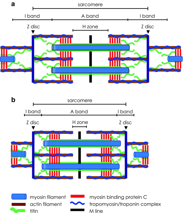
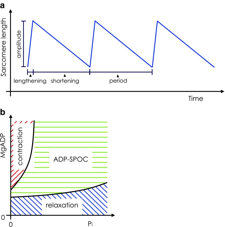
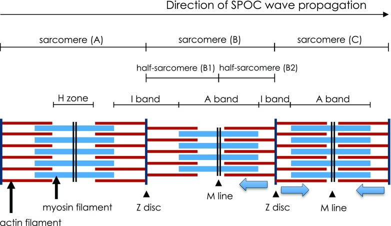
Striated muscle is well known to exist in either of two states-contraction or relaxation-under the regulation of Ca2+ concentration. Described here is a less well-known third, intermediate state induced under conditions of partial activation, known as SPOC (SPontaneous Oscillatory Contraction). This state is characterised by auto-oscillation between rapid-lengthening and slow-shortening phases. Notably, SPOC occurs in skinned muscle fibres and is therefore not the result of fluctuating Ca2+ levels, but is rather an intrinsic and fundamental phenomenon of the actomyosin motor. Summarised in this review are the experimental data on SPOC and its fundamental mechanism. SPOC presents a novel technique for studying independent communication and coordination between sarcomeres. In cardiac muscle, this auto-oscillatory property may work in concert with electro-chemical signalling to coordinate the heartbeat. Further, SPOC may represent a new way of demonstrating functional defects of sarcomeres in human heart failure.

Keywords: Auto-oscillation; Cardiac muscle; SPOC; Sarcomere; Skeletal muscle.


Figures

Fig. 1
Fig. 1
Structural and contractile proteins of the striated muscle sarcomere in relaxed (a) and contracted (b) states. a The sarcomere extends between two adjacent Z discs. The Z disc anchors the actin filaments with their overlying tropomyosin/troponin complex. The M line defines the centre of the sarcomere, bisects the myosin filaments and extends across the width of the A band. The I band is the gap between the free ends of adjacent thick filaments in the A band and is bisected by the Z disc. A giant elastic protein, titin, extends from the Z disc through the I band, attaches to the thick filaments of the A band and ends at the M line. Myosin binding protein C (MyBP-C) is located in the part of the A band where cross-bridges are formed. b Graphic representation of how contraction occurs by mutual sliding of the thick and thin filaments while the lengths of the filaments remain unchanged. Thus, during sarcomere shortening, both the H zone and I band progressively narrow while there is no change in the A band
Fig. 2
Fig. 2
a Schematic of oscillations in sarcomere length during SPontaneous Oscillatory Contraction (SPOC). The total SPOC period comprises rapid-lengthening and slow-shortening phases. b Two-dimensional state-diagram of striated muscle showing contraction, relaxation and ADP–SPOC plotted against inorganic phosphate (Pi; x-axis) and MgADP (y-axis) concentrations, where MgATP concentration is fixed. SPOC requires the coexistence of MgATP and MgADP above a certain threshold proportion. Therefore, if the fixed concentration of MgATP is increased, a higher MgADP concentration is required to induce SPOC. Adapted from Shimizu et al. (1992)
Fig. 3
Fig. 3
An illustration of three adjacent sarcomeres (A, B and C) during SPOC. As the SPOC wave progresses from left to right, sarcomere A is in the lengthening phase, and sarcomere C is in the shortening phase. Sarcomere B is at the interface between phases and demonstrates asymmetric length increase in half-sarcomere B1, while half-sarcomere B2 remains in the shortening phase. Lengthening in half-sarcomere B1 results in a quick release of tension at adjacent half-sarcomere B2. Half-sarcomere B1 redevelops tension soon after lengthening, causing half-sarcomere B2 to lengthen under the newly imposed strain. In this way, the SPOC wave propagates along successive half-sarcomeres down the length of the myofibril. Adapted from Ishiwata et al. (1991)
Fig. 4
Fig. 4
Relationship between ADP–SPOC and resting heart rate. a Period of ADP–SPOC correlated with the period of resting heart rate across animal species (correlation coefficient 0.976), b shortening velocity of SPOC very closely correlated with resting heart rate (correlation coefficient 0.990), with a near-proportional relationship (bpm beats per minute), c Strong correlation between shortening velocity during ADP–SPOC and the sliding velocity of actin filaments determined by in vitro motility assay (correlation coefficient 0.987), though the relationship was not a proportional one. Data are presented as the mean ± standard deviation (SD). Solid lines Regression lines, broken lines regression lines that cross the origin, indicating a proportional relationship. Graphs adapted from Sasaki et al. (2005)

Similar articles

Cited by

References

    1. Abdulla R, Blew GA, Holterman MJ. Cardiovascular embryology. Pediatr Cardiol. 2004;25:191–200. - PubMed
    1. Alcalai R, Seidman JG, Seidman CE. Genetic basis of hypertrophic cardiomyopathy: From bench to the clinics. J Cardiovasc Electrophysiol. 2008;19:104–110. - PubMed
    1. Anazawa T, Yasuda K, Ishiwata S. Spontaneous oscillation of tension and sarcomere length in skeletal myofibrils. Microscopic measurement and analysis. Biophys J. 1992;61:1099–1108. doi: 10.1016/S0006-3495(92)81919-7. - DOI - PMC - PubMed
    1. Dillmann WH. Hormonal influences on cardiac myosin ATPase activity and myosin isoenzyme distribution. Mol Cell Endocrinol. 1984;34:169–181. doi: 10.1016/0303-7207(84)90173-4. - DOI - PubMed
    1. Ebashi S, Endo M. Calcium ions and muscle contraction. Prog Biophys Mol Biol. 1968;18:123–183. doi: 10.1016/0079-6107(68)90023-0. - DOI - PubMed

LinkOut - more resources