Review
. 2016 Dec;8(4):409-427.
doi: 10.1007/s12551-016-0218-6. Epub 2016 Oct 6.

Dynamic light scattering: a practical guide and applications in biomedical sciences

Affiliations
Review

Dynamic light scattering: a practical guide and applications in biomedical sciences

Jörg Stetefeld et al. Biophys Rev. 2016 Dec.

Abstract

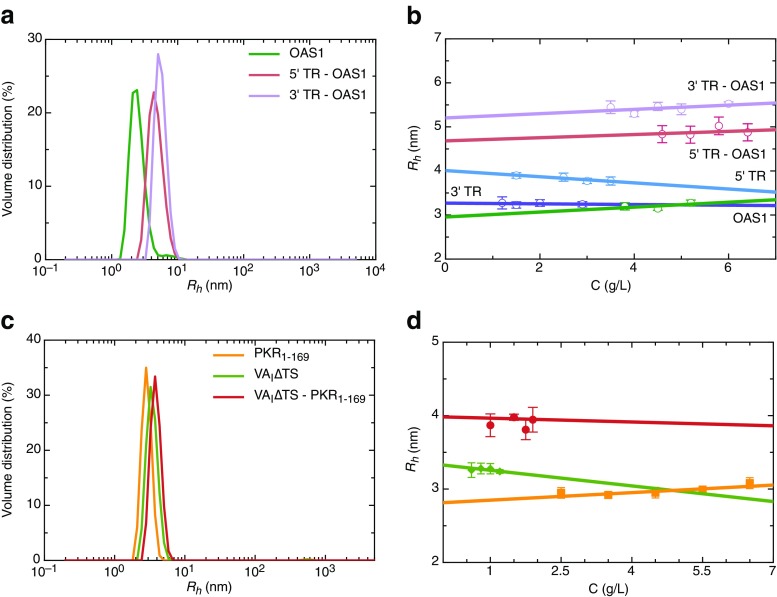
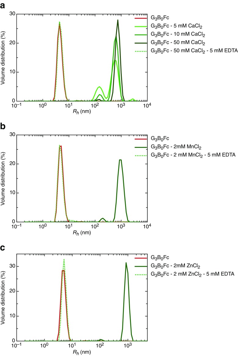
Dynamic light scattering (DLS), also known as photon correlation spectroscopy (PCS), is a very powerful tool for studying the diffusion behaviour of macromolecules in solution. The diffusion coefficient, and hence the hydrodynamic radii calculated from it, depends on the size and shape of macromolecules. In this review, we provide evidence of the usefulness of DLS to study the homogeneity of proteins, nucleic acids, and complexes of protein-protein or protein-nucleic acid preparations, as well as to study protein-small molecule interactions. Further, we provide examples of DLS's application both as a complementary method to analytical ultracentrifugation studies and as a screening tool to validate solution scattering models using determined hydrodynamic radii.

Keywords: Analytical ultracentrifuge; Diffusion coefficient; Dynamic light scattering; Hydrodynamic radius; Light scattering; Protein–ligand interactions; Protein–nucleic acid complexes; Protein–protein complexes.


Conflict of interest statement

Conflict of Interests

Jörg Stetefeld declares that he has no conflicts of interest. Sean A. McKenna declares that he has no conflicts of interest. Trushar R. Patel declares that he has no conflicts of interest.

Ethical approval

This article does not contain any studies with human participants or animals performed by any of the authors.

Figures

Fig. 1
Fig. 1
Evaluating homogeneity of protein solution. a Hydrodynamic radius distribution by intensity for nidogen-1 (ND-1) at 0.4 mg/ml, indicating that although there is a major peak for each measurement, there are multiple peaks visible between 15 nm and 500 nm (x-axis) suggesting the presence of high molecular weight aggregated species. Degradation at 4 nm is also visible for one of the measurements in red colour. b Hydrodynamic radius distribution by volume derived from intensity profiles for ND-1 at 0.4 mg/ml concentration, suggesting that volume distribution is not sensitive enough for detecting scant amount of aggregation. However, peaks from multiple runs do not coincide with each other: that essentially is a reflection of heterogeneous sample. Again, the degradation is visible for the measurement that is clearly visible for intensity distribution. c Similar to a, the aggregation studies for ND-1 were performed at 0.5 mg/m for a preparation optimized to obtain cleaner sample, indicating that compared to a, the level of aggregation is severely reduced. d Homogeneity of ND-1 reflected by volume distribution of nearly identical peaks from multiple measurements, demonstrating highly purified and homogenous preparation. Recombinant ND-1 (139 kDa) was expressed into 293-EBNA cells and purified using affinity chromatography, followed by SEC in 50 mM Tris–HCl (pH 7.5), 200 mM NaCl buffer as described earlier (Patel et al. 2014)
Fig. 2
Fig. 2
Homogeneity studies of ovalbumin. a and b represents R h distribution at multiples concentrations for ovalbumin. The intensity distributions (a) suggest presence of ∼5 % aggregated material at 100 nm and above. Those peaks are not present in volume distribution profiles (b) since the amount of aggregated material is negligible. However, since large molecules scatter light more strongly, even a trace amount of aggregation can be detected using intensity distribution. In both cases, nearly identical positions of peak suggest a homogenous preparation. For biophysical and structure biology applications, such preparations are acceptable; however, for therapeutic antibodies, further optimization may be required. c Plot of R h vs concentration for ovalbulin, where the y-axis intercept provides concentration-independent R h of 2.88 ± 0.06 nm. d Hydrodynamic radius for frequently used molecular weight standards to calibrate size exclusion column was measured at single concentration. The details for molecular weights and concentrations are provided in Table 2. Note that all of the proteins used here are globular proteins; therefore, it is possible to compare their R h profiles in terms of molecular weight. The smallest protein here is aprotinin (Mw 6.5 kDa) with R h of 1.82 nm, a value that increases with increase in molecular weight to 8.71 nm for thyroglobulin (Mw 670 kDa). Ovalbumin (44 kDa) was purchased from Fischer Scientific (BP2535) and solubilised using 20 mM phosphate buffer (pH 7.0) for DLS measurements (Scott et al. 2011). The molecular weight standards kit was purchased from GE healthcare, and DLS data were collected in 50 mM Tris (pH 8.0), 150 mM NaCl buffer
Fig. 3
Fig. 3
Homogeneity of protein-protein complexes. a Hydrodynamic radius distribution of ND-1, laminin γ-1 (LM γ−1) arm, and their complex at 3 mg/ml indicating purity of individual species as well as their complex. b Concentration dependence of ND-1, LM γ−1 arm, and their complex up to 4 mg/ml (Patel et al. 2014). Recombinant ND-1 (139 kDa) and LM γ-1 (109 kDa) were expressed into 293-EBNA cells and purified using affinity chromatography followed by SEC. Complex of ND-1-LM γ-1 was purified using SEC, and DLS measurements were performed in 50 mM Tris–HCl (pH 7.5), 200 mM NaCl buffer as described earlier (Patel et al. 2014)
Fig. 4
Fig. 4
Evaluating homogeneity of viral RNA preparations. Multiple measurements for intensity distribution (a) and volume distribution (b) at 3.5 mg/ml suggest that the 5’ TR RNA is highly pure and devoid of any aggregation. Similarly, 3’ untranslated region (3’ TR) from West Nile virus was also studied using DLS at 2.7 mg/ml, where a and b present intensity and volume distributions suggesting presence of a homogenous solution of 3’ TR. The 5’ and 3’ untranslated regions (5’ TR — 47.5 kDa, 3’ TR — 37.5 kDa) from West Nile virus were prepared using an in-vitro transcription method followed by purification using SEC and DLS analysis in 50 mM Tris (pH 7.0), 100 mM NaCl buffer as described earlier (Deo et al. 2014, 2015)
Fig. 5
Fig. 5
Homogeneity of protein-RNA complexes. a Homogeneity studies of OAS1 (2.2 mg/ml), 5’ TR–OAS1 complex (4.0 mg/ml), and 3’ TR–OAS1 complex (3.5 mg/ml). b The concentration dependence of OAS1, 5’ TR, 3’ TR, 5’ TR - OAS1, and 3’ TR–OAS1 complex. c Hydrodynamic radius distribution for PKR1–169 (3.5 mg/ml), VAI∆TS (1.2 mg/ml), and VAI∆TS–PKR1–169 complex (1 mg/ml), suggesting homogeneity of all three species. d Dynamic light scattering data for PKR1–169, VAI∆TS, and VAI∆TS–PKR1–169 complex are presented at multiple concentrations. Human OAS1 (41 kDa) was expressed in BL21(DE3) cells followed by purification by means of affinity and SEC (Meng et al. 2012). DLS data were measured in 50 mM Tris (pH 7.5), 100 mM NaCl, 1 mM DTT buffer (Deo et al. 2014). The 5’ TR–OAS1 and 3’–TR OAS1 complexes were purified using SEC and analysed using DLS in 50 mM Tris (pH 7.5), 40 mM NaCl, and 1 mM EDTA (Deo et al. 2015). Similarly, the adenovirus VAI RNA lacking the terminal stem-loop RNA (VAI∆TS, 32.3 kDa) was prepared by in-vitro transcription method followed by purification using SEC (Dzananovic et al. 2014), whereas human protein kinase R (PKR) lacking the C-terminal kinase domain (PKR1–169 18.8 kDa) was expressed into BL21(DE3) cells, purified by means of affinity and SEC followed by DLS data collection in 50 mM Tris (pH 7.50), 100 mM NaCl, and 5 mM 2-mercaptoethanol (Dzananovic et al. 2013). DLS data for SEC purified VAI∆TS–PKR1–169 complex was collected in 50 mM Tris (pH 7.50), 100 mM NaCl buffer (Dzananovic et al. 2014)
Fig. 6
Fig. 6
Protein–small molecules interaction studies. The mouse G3B0 protein fused with human Fc domain of IgG–G3B0Fc was studied using DLS at 1 mg/ml. a, b, and c presents volume distribution profiles for G3B0Fc protein and its interactions with divalent cations. a A single peak for G3B0Fc appears at ∼5 nm which resolves into two peaks at ∼ 100 nm and ∼ 600 nm upon addition of 5 mM CaCl2. The higher molecular weight peak further increases up to ∼ 700 nm in the presence of 50 mM CaCl2. However, addition of EDTA (5 mM) reduces the huge complex of G3B0Fc–50 mM CaCl2 to ∼ 5 nm that essentially overlays on G3B0Fc data. Similarly, addition of only 2 mM MnCl2 (b) and 2 mM ZnCl2 (c) increases the size of G3B0Fc from ∼ 5 nm to ∼900 nm, indicating presence of a very heavy aggregate. Addition of EDTA (5 mM in both cases) again reduces the heavy complex to monomeric size of G3B0Fc. The C-terminal laminin G-like G3 domain variant of chicken agrin without insert of amino acids at the alternative splice site B (B0), G3B0 was expressed as a fusion protein, with human Fc domain of IgG–G3B0Fc being expressed using HEK293 cells as described previously (Patel et al. 2011). The G3B0Fc protein was affinity purified, followed by DLS data collection in 20 mM Tris, 150 mM NaCl at pH 7.2

Similar articles

Cited by

References

    1. Amoros D, Ortega A, de la Torre JG. Hydrodynamic properties of wormlike macromolecules: Monte Carlo simulation and global analysis of experimental data. Macromolecules. 2011;44:5788–5797. doi: 10.1021/ma102697q. - DOI
    1. Ariyo EO, et al. Biophysical Characterization of G-Quadruplex Recognition in the PITX1 mRNA by the Specificity Domain of the Helicase RHAU. PLoS One. 2015;10:e0144510. doi: 10.1371/journal.pone.0144510. - DOI - PMC - PubMed
    1. Barnett CE. Some applications of wave-length turbidimetry in the infrared. J Phys Chem. 1942;46:69–75. doi: 10.1021/j150415a009. - DOI
    1. Benitez AA, Hernandez Cifre JG, Diaz Banos FG, de la Torre JG. Prediction of solution properties and dynamics of RNAs by means of Brownian dynamics simulation of coarse-grained models: Ribosomal 5S RNA and phenylalanine transfer RNA. BMC Biophys. 2015;8:11. doi: 10.1186/s13628-015-0025-7. - DOI - PMC - PubMed
    1. Berne BJ, Pecora R. Dynamic light scattering: with applications to chemistry, biology, and physics. New York, USA: John Wiley & Sons, Inc; 1976.