Review
. 2016 Dec;8(4):385-396.
doi: 10.1007/s12551-016-0230-x. Epub 2016 Nov 5.

Molecular assembly and structure of the bacteriophage T4 tail

Affiliations
Review

Molecular assembly and structure of the bacteriophage T4 tail

Fumio Arisaka et al. Biophys Rev. 2016 Dec.

Abstract

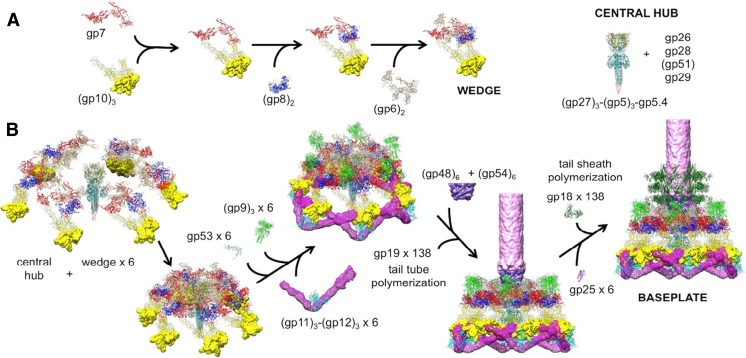
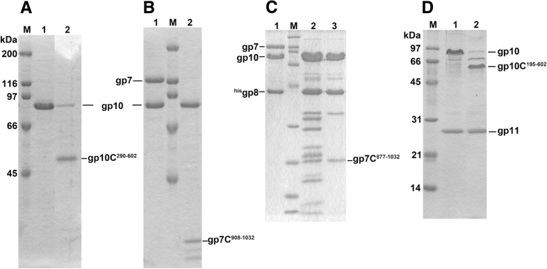
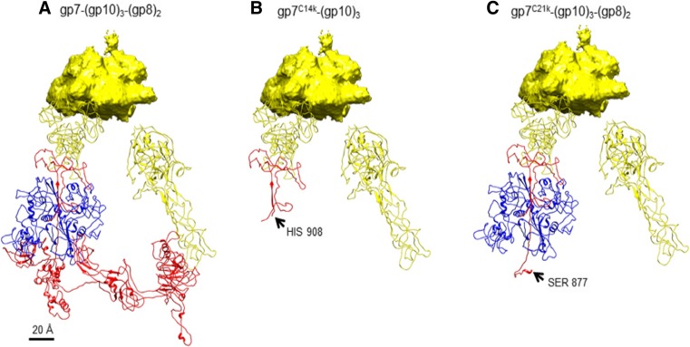
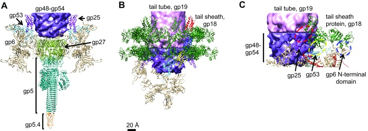
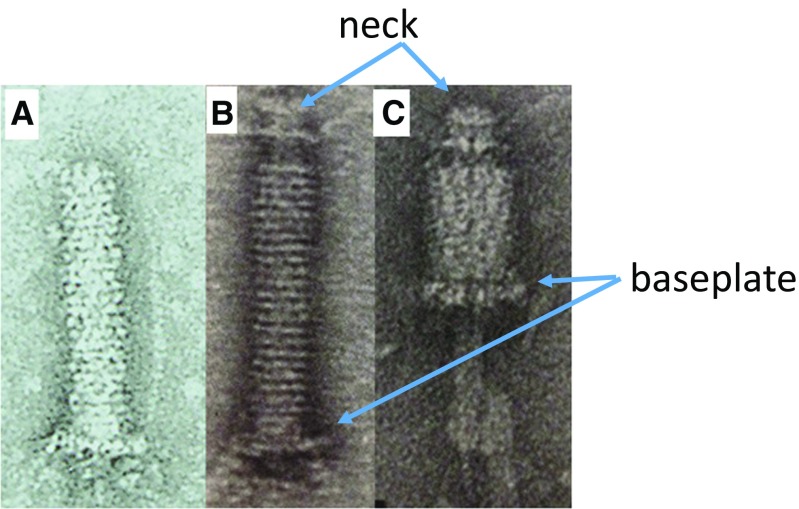
The tail of bacteriophage T4 undergoes large structural changes upon infection while delivering the phage genome into the host cell. The baseplate is located at the distal end of the contractile tail and plays a central role in transmitting the signal to the tail sheath that the tailfibers have been adsorbed by a host bacterium. This then triggers the sheath contraction. In order to understand the mechanism of assembly and conformational changes of the baseplate upon infection, we have determined the structure of an in vitro assembled baseplate through the three-dimensional reconstruction of cryo-electron microscopy images to a resolution of 3.8 Å from electron micrographs. The atomic structure was fitted to the baseplate structure before and after sheath contraction in order to elucidate the conformational changes that occur after bacteriophage T4 has attached itself to a cell surface. The structure was also used to investigate the protease digestion of the assembly intermediates and the mutation sites of the tail genes, resulting in a number of phenotypes.

Keywords: Assembly; Bacteriophage; Contractile tail; Infection; Molecular recognition; Tail baseplate.


Conflict of interest statement

Conflict of interest

Fumio Arisaka declares that none of the authors have any conflicts of interest.

Ethical approval

This article does not contain any studies with human participants or animals performed by the author.

Figures

Fig. 1
Fig. 1
Assembly of a baseplate based on present and earlier results. a Wedge assembly. Gp10, gp8 and gp6 bind sequentially to the gp7 backbone protein. The central hub of the baseplate is assembled independently. b Baseplate and tail assembly. Six wedges assemble around the central hub to form a baseplate. Gp53 binds adjacent wedges together, and gp9 and the gp11–gp12 complex subsequently bind to the baseplate, further stabilizing the dome-shaped configuration. Then, gp48 and gp54 bind to the top of the central hub and initiate polymerization of the tail tube. Gp25 attaches to the gp48–gp54 complex, initiating polymerization of the tail sheath. For clarity, only three rings of the tail sheath are shown (adapted from Fig. 4 of Yap et al. 2016). gp Gene product
Fig. 2
Fig. 2
Lysylendopeptidase digestion of the wedge intermediates. a (gp10)3 before (lane 1) and after (lane 2) digestion. b (gp7)(gp10)3 before (lane 1) and after (lane 2) digestion. In the presence of gp7, gp10 becomes protease resistant and binds the 14-kDa C-terminal fragment of gp7 (gp7C residues 908–1032). c (gp7)(gp10)3(gp8)2 before (lane 1) and after (lane 2) digestion; lane 3 Size Exclusion Chromatography purified digested product. In the presence of gp8, the associated C-terminal fragment of gp7 to gp10 becomes larger to be 18 kDa (gp7C residues 877–1032). d (gp10)3(gp11)3 before (lane 1) and after (lane 2) digestion. In the presence of gp11, gp10 becomes partially protease resistant and gives larger C-terminal fragment (gp10C residues 196–602). The rationale of this difference is now explained based on the quaternary structure of the complex and discussed in the text. Lane M Molecular weight marker. In a and d, the molecular weights are indicated in the figure; in b, the molecular weights are the same as in a; in c, the molecular weights are 200, 116, 97, 66, 45, 31, 21, 14 and 6 kDa, respectively, beginning from the top of the gel
Fig. 3
Fig. 3
a The (gp10)3(gp7)(gp8)2 complex in the in vitro-assembled baseplate complex. b The (gp10)3(gp7) complex after lysylendopeptidase cleavage. c The (gp10)3(gp7)(gp8)2 complex after digestion by lysylendopeptidase. Colors: yellow:gp10, red:gp7, blue:gp8.
Fig. 4
Fig. 4
Baseplate completion and sheath formation. a Interaction between gp6 and gp27, a hub protein. b Formation of the first and second annuli of gp18.The C-terminal structure of gp18 which resembles gp25 is indicated by red. c Interactions between gp18 and the baseplate proteins gp6, gp53 and gp25. Tube polymerization starts with (gp48)6(gp54)6 binding. Then after gp25 binds, tail sheath association or polymerization will start. C-terminal structure of gp18 which resembles gp25 is shown in red
Fig. 5
Fig. 5
Tails (a), tails isolated from 23 infections (b) and “necked tail” (c) Images of contracted “necked tail” (b, c) from Coombs and Arisaka 1994)
Fig. 6
Fig. 6
Crystal structure of gp18 (residues 20–510) (Aksyuk et al. 2009) in combination with the predicted C-terminal domain (Fokine et al. 2013). Four domains of gp18 are shown in blue (Domain I), olive green (Domain II), orange–red (Domain III) and green (C-terminal domain). The N-terminal 20 residues and residues 484–496 are not ordered in the crystal structure and thus not observed. Residue 510 is the last residue in the crystal structure. Mutation sites with a number of phenotypes were mapped by DNA sequencing of the mutants (Takeda et al. 2004). The temperature-sensitive mutants (ts; grow at 30 °C, but not at 42 °C) were mapped as W34R (or P), Q39P, P219L, G257D, Q277Y (or C, R, F), W487R, G587N and Q589Y. The heat-sensitive mutants (hs; lose viability upon incubation for 30 min at 55 °C) were mapped as P5S, R565C and G598S. The cold-sensitive mutants (cs; grow at 37 °C, but not at 25 °C) were mapped as R584H, Q589E and Q592E (or R); they are in the C-terminal domain circled). The carbowax mutants (CBW; can infect in the presence of a high concentration of polyethylene glycol) were mapped at G106S, S175F and A178V, in Domain I, and the mutation sites are circled
Fig. 7
Fig. 7
Schematic map of amino acid replacements and phenotypes on the primary structure of gp18, the tail sheath protein. Black and white lines correspond to protease-resistant domain and PS17 phage sheath-homologous regions, respectively. (Takeda et al. 2004). See section Tail sheath protein gp18 for definition of mutations
Fig. 8
Fig. 8
a. Structure of (gp27)3(gp5)3(gp5.4)1 to show the location of the pseudo-sixfold symmetry of gp27 and pseudo threefold symmetry of gp5.4. b. Four domains of gp27, where Domains I and III constitute part of the pseudo sixfold symmetry as shown in c. c Overview of gp27 and gp5.4 to show the pseudo sixfold symmetry of (gp27)3 and pseudo threefold symmetry of gp5.4. d. Parallel β-sheets formed between gp5.4 and gp5C. #1, #2, #3 Three identical subunits (numbers are arbitrary)
Fig. 9
Fig. 9
Crystal structure of gp5 structure (residues 1–575) (Kanamaru et al. 2002). Only one polypeptide chain of the trimer is shown for clarity. ts and hs mutation sites were mapped by DNA sequencing of the mutants, with ts mutants mapped as A65T, T80A, A276T, G322D(5ts1) and E337K, and hs mutants mapped as P122S and A352T (Takeda et al. 1998). See section Tail sheath protein gp18 for definition of mutations
Fig. 10
Fig. 10
Schematic map of amino acid replacements and phenotypes on the primary structure of gp5, the tail lysozyme protein. Black region of bar corresponds to T4-lysozyme homologous region. Five ts mutation sites and two hs sites as well as three amber sites are listed (Takeda et al. 1998). See section Tail sheath protein gp18 for definition of mutations
Fig. 11
Fig. 11
Structure of gp15-18 before (a, c) and after (b, d) after sheath contraction) and schematic longitudinal cross-section of the neck area (adapted from Figs. 1 c, d and 6 c–f by Fokine et al. 2013)

References

    1. Abuladze NK, Gingery M, Tsai I, Eiserling FA. Tail length determination in bacteriophage T4. Virology. 1994;199:301–310. doi: 10.1006/viro.1994.1128. - DOI - PubMed
    1. Akhter T, Zhao L, Kohda A, Mio K, Kanamaru S, Arisaka F. The neck of bacteriophage T4 is a ring-like structure formed by a hetero-oligomer of gp13 and gp14. Biochim Biophys Acta. 2007;1774(8):1036–1043. doi: 10.1016/j.bbapap.2007.05.011. - DOI - PubMed
    1. Aksyuk AA, Leiman PG, Kurochkina LP, Schneider MM, Kostyuchenko VA, Mesyanzhinov VV, Rossmann MG. The tail sheath structure of bacteriophage T4: a molecular machine for infecting bacteria. EMBO J. 2009;28:821–829. doi: 10.1038/emboj.2009.36. - DOI - PMC - PubMed
    1. Arisaka F, Tschopp J, van Driel R, Engel J. Reassembly of the bacteriophage T4 tail from the core­baseplate and the monomeric sheath protein P18: a cooperative association process. J Mol Biol. 1979;132:369–386. doi: 10.1016/0022-2836(79)90266-3. - DOI - PubMed
    1. Arisaka F, Engel J, Klump J. Contraction and dissociation of the bacteriophage T4 tail sheath induced by heat and urea. In: DuBow M, editor. Bacteriophage assembly. New York: Alan R. Liss; 1981. pp. 365–379. - PubMed

LinkOut - more resources