Review
. 2010 Dec;2(4):181-189.
doi: 10.1007/s12551-010-0040-5. Epub 2010 Nov 20.

Synaptopodin family of natively unfolded, actin binding proteins: physical properties and potential biological functions

Affiliations
Review

Synaptopodin family of natively unfolded, actin binding proteins: physical properties and potential biological functions

Joseph M Chalovich et al. Biophys Rev. 2010 Dec.

Abstract

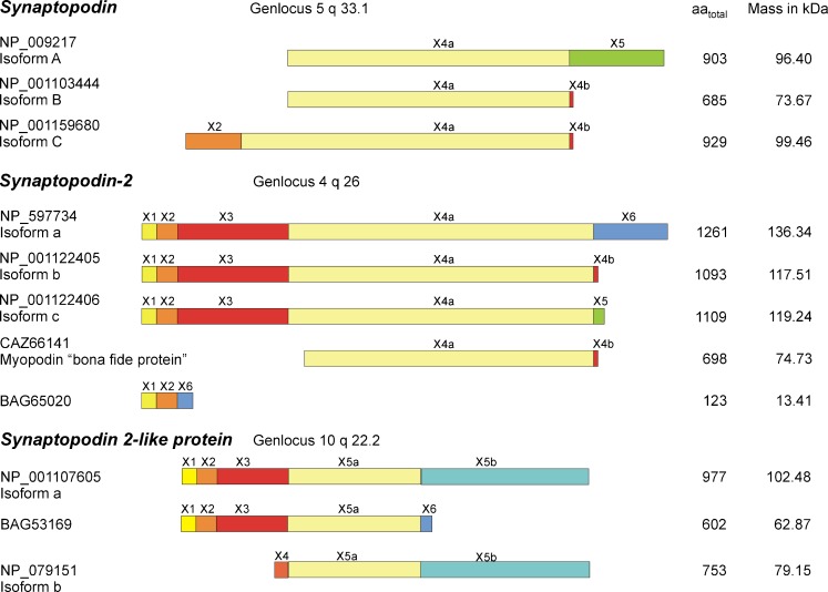
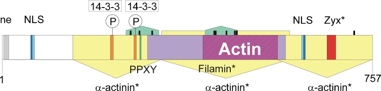
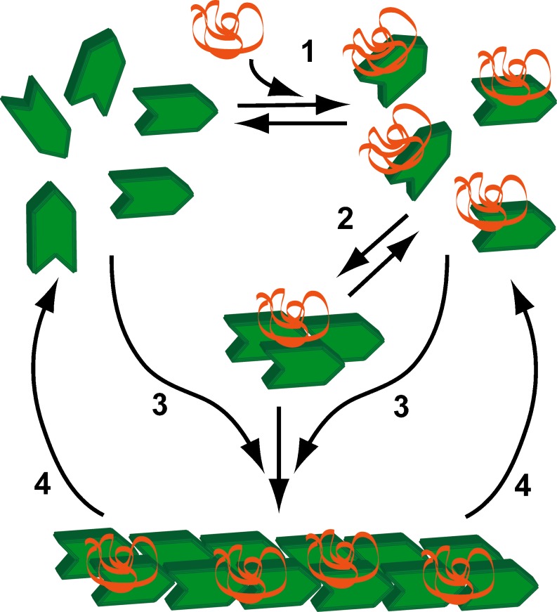
The synaptopodin family of proteins consists of at least 3 members: synaptopodin, the synaptopodin 2 proteins, and the synaptopodin 2-like proteins. Each family member has at least 3 isoforms that are produced by alternative splicing. Synaptopodin family members are basic proteins that are rich in proline and have little regular 2° or 3° structure at physiological temperature, pH and ionic strength. Like other natively unfolded proteins, synaptopodin family members have multiple binding partners including actin and other actin-binding proteins. Several members of the synaptopodin family have been shown to stimulate actin polymerization and to bundle actin filaments either on their own or in collaboration with other proteins. Synaptopodin 2 has been shown to accelerate nucleation of actin filament formation and to induce actin bundling. The actin polymerization activity is inhibited by Ca2+-calmodulin. Synaptopodin 2 proteins are localized in Z-bands of striated and heart muscle and dense bodies of smooth muscle cells. Depending on the developmental status and stress, at least one member of the synaptopodin family can occupy nuclei of some cells. Members of the synaptopodin 2 subfamily have been implicated in cancers.

Keywords: Actin bundling; Actin polymerization; Cancer; Natively unfolded; Nuclear transport; Synaptopodin.


Figures

Fig. 1
Fig. 1
Primary protein structure of synaptopodin family members and their splice forms. Each of the three members of the synaptopodin family can exist in at least three different isoforms produced by alternative splicing of the N- and C-terminal exons. The figure shows only the composition of the proteins, therefore non-coding exons 1 and 3 in synaptopodin are not shown. Except for the unnamed protein that is listed with the NCBI accession number BAG65020 the isoforms of each subfamily share a common core region
Fig. 2
Fig. 2
Binding motifs in mouse myopodin CAC67798. This region forms the core region of synaptopodin 2 encoded by exon 4a. Motifs that have only been described in human myopodin are marked with a star. The green domains represent proline-rich stretches. The heavy black bars are PXXP motifs that are putative SH3 interacting domains. The yellow arrows show three independent α-actinin binding regions (Linnemann et al. 2010). The filamin binding domain (Linnemann et al. 2010), shown by the light violet bar contains the main actin binding region (Weins et al. 2001) (dark purple diagonals). The region marked ne has weak nuclear export activity without a classical NES-motif (Van Impe et al. 2003). NLS is the nuclear localization site; the consensus motifs are in dark blue while additional motifs (De Ganck et al. 2005) are in light blue. Binding motifs for 14-3-3 are shown along with the phosphorylation sites S225 and T272, that are targets of protein kinase A and calcium-calmodulin kinase II (Faul et al. 2005, 2007). The PPXY motif may interact with WW-domain containing proteins. Zyx is the Zyxin binding motif discovered in human myopodin (Yu and Luo 2006)
Fig. 3
Fig. 3
Proposed effect of synaptopodin 2 on actin polymerization. 1 G-actin (green) binds to synaptopodin 2 (orange ribbon). 2 Actin-synaptopodin 2 complexes rapidly form nuclei consisting of 1 synaptopodin 2 and 3 actin monomers. 3 Elongation of synaptopodin 2 nuclei occurs rapidly; G-actin-synaptopodin 2 complexes add more rapidly than plain G-actin. 4 Actin can detach from both the barbed (fast growing) and pointed ends of actin filaments. The primary effect of Ca2+-calmodulin appears to be to reduce the rate of nucleation (step 2)

References

    1. Albiges-Rizo C, Destaing O, Fourcade B, Planus E, Block MR. Actin machinery and mechanosensitivity in invadopodia, podosomes and focal adhesions. J Cell Sci. 2009;122:3037–3049. doi: 10.1242/jcs.052704. - DOI - PMC - PubMed
    1. Asanuma K, Kim K, Oh J, Giardino L, Chabanis S, Faul C, Reiser J, Mundel P. Synaptopodin regulates the actin-bundling activity of alpha-actinin in an isoform-specific manner. J Clin Invest. 2005;115:1188–1198. - PMC - PubMed
    1. Asanuma K, Yanagida-Asanuma E, Faul C, Tomino Y, Kim K, Mundel P. Synaptopodin orchestrates actin organization and cell motility via regulation of RhoA signalling. Nat Cell Biol. 2006;8:485–491. doi: 10.1038/ncb1400. - DOI - PubMed
    1. Beall B, Chalovich JM. Fesselin, a synaptopodin-like protein, stimulates actin nucleation and polymerization. Biochemistry. 2001;40:14252–14259. doi: 10.1021/bi011806u. - DOI - PMC - PubMed
    1. Beqqali A, Monshouwer-Kloots J, Monteiro R, Welling M, Bakkers J, Ehler E, Verkleij A, Mummery C, Passier R. CHAP is a newly identified Z-disc protein essential for heart and skeletal muscle function. J Cell Sci. 2010;123:1141–1150. doi: 10.1242/jcs.063859. - DOI - PubMed

LinkOut - more resources