Review
. 2016 Jun;8(2):179-191.
doi: 10.1007/s12551-016-0199-5. Epub 2016 Apr 30.

The two sides of a lipid-protein story

Affiliations
Review

The two sides of a lipid-protein story

Luis G Mansor Basso et al. Biophys Rev. 2016 Jun.

Abstract

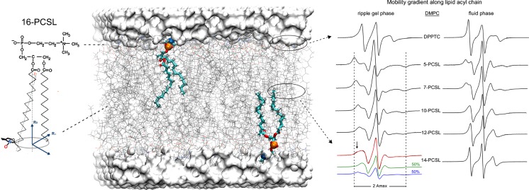
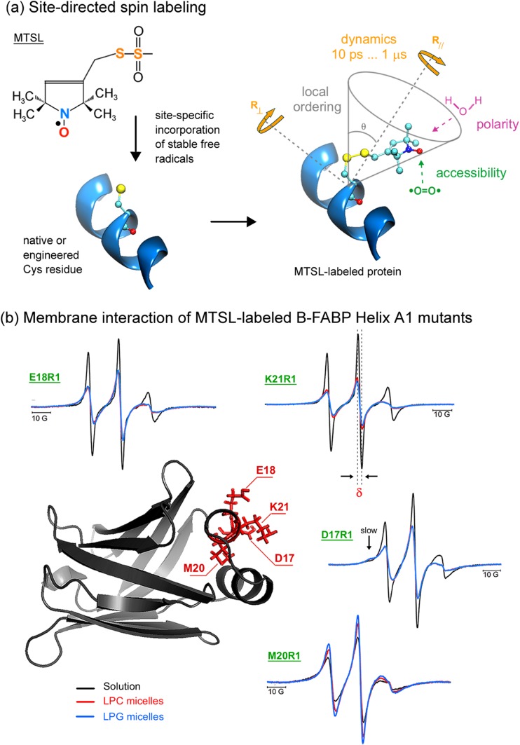
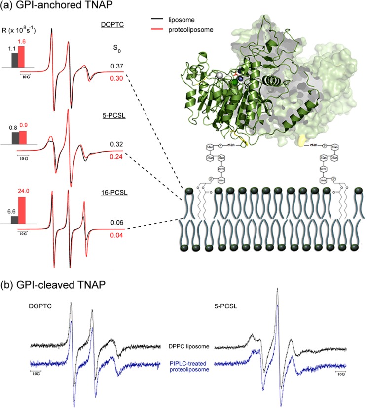
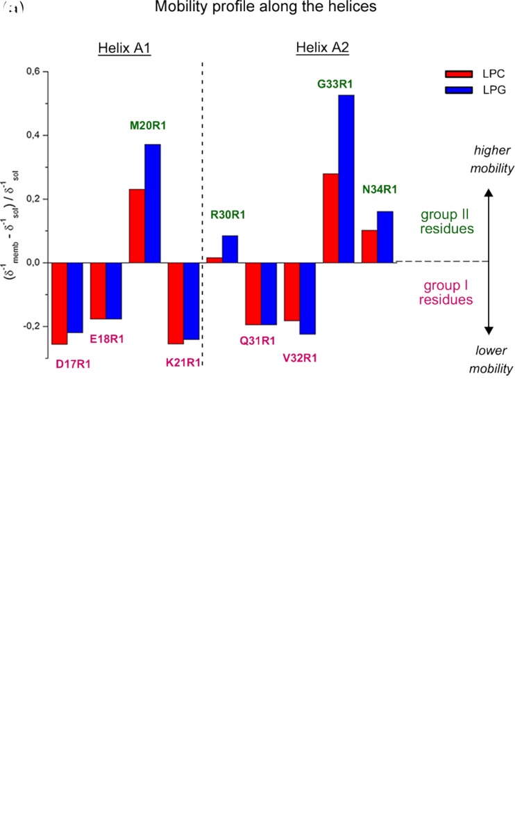
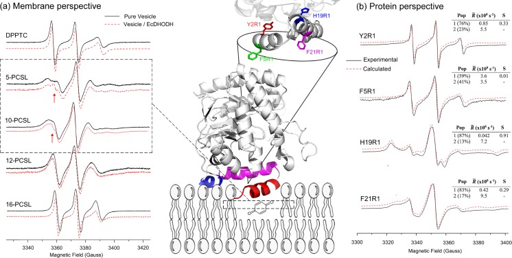
Protein-membrane interactions play essential roles in a variety of cell functions such as signaling, membrane trafficking, and transport. Membrane-recruited cytosolic proteins that interact transiently and interfacially with lipid bilayers perform several of those functions. Experimental techniques capable of probing changes on the structural dynamics of this weak association are surprisingly limited. Among such techniques, electron spin resonance (ESR) has the enormous advantage of providing valuable local information from both membrane and protein perspectives by using intrinsic paramagnetic probes in metalloproteins or by attaching nitroxide spin labels to proteins and lipids. In this review, we discuss the power of ESR to unravel relevant structural and functional details of lipid-peripheral membrane protein interactions with special emphasis on local changes of specific regions of the protein and/or the lipids. First, we show how ESR can be used to investigate the direct interaction between a protein and a particular lipid, illustrating the case of lipid binding into a hydrophobic pocket of chlorocatechol 1,2-dioxygenase, a non-heme iron enzyme responsible for catabolism of aromatic compounds that are industrially released in the environment. In the second case, we show the effects of GPI-anchored tissue-nonspecific alkaline phosphatase, a protein that plays a crucial role in skeletal mineralization, and on the ordering and dynamics of lipid acyl chains. Then, switching to the protein perspective, we analyze the interaction with model membranes of the brain fatty acid binding protein, the major actor in the reversible binding and transport of hydrophobic ligands such as long-chain, saturated, or unsaturated fatty acids. Finally, we conclude by discussing how both lipid and protein views can be associated to address a common question regarding the molecular mechanism by which dihydroorotate dehydrogenase, an essential enzyme for the de novo synthesis of pyrimidine nucleotides, and how it fishes out membrane-embedded quinones to perform its function.

Keywords: EPR; ESR; Metalloproteins; Protein–lipid interaction; Protein–membrane interaction; Spin labeling.


Conflict of interest statement

Conflict of Interest

Luis G. Mansor Basso declares that he has no conflict of interest.

Luis F. Santos Mendes declares that he has no conflict of interest.

Antonio J. Costa-Filho declares that he has no conflict of interest.

Ethical approval

This article does not contain any studies with human participants or animals performed by any of the authors.

Figures

Fig. 1
Fig. 1
Spin labeling ESR from the membrane perspective and structural dynamics-line-shape correlations. a Dimiristoylphosphatidylcholine (DMPC) lipid bilayer (stick representation in gray) usually doped with 0.5–1.0 mol% of spin-labeled lipids containing a nitroxide radical attached to different positions along the lipid acyl chain, such as the spin label 16-PCSL on the left. A particular phospholipid is highlighted in licorice representation with the phosphorus and nitrogen atoms of the lipid head group colored in orange and blue, respectively. Different n-PCSL (n = 5, 7, 10, 12, 14, and 16) spin labels report on specific regions of the lipid bilayer. On the right, typical n-PCSL ESR spectra obtained for DMPC in the ripple gel phase (20 °C) and in fluid phase (35 °C). It is worth noting the lineshape changes of the spectra due to the mobility gradient experienced by the spin labels from the head group region down to the hydrophobic core of the lipid bilayer and also the lipid phase-dependence of the lineshape. Coexistence of two spin populations presenting different ordering and dynamics can also be detected by ESR, as shown by the 14-PCSL in the DMPC ripple gel phase. The lipid bilayer was built with CHARMM-GUI Membrane Builder (http://www.charmm-gui.org/input/membrane) (Jo et al. ; Wu et al. 2014) and rendered with Visual Molecular Dynamics (Humphrey et al. 1996). Adapted from (Basso et al. 2011) with permission
Fig. 2
Fig. 2
Spin labeling ESR from the protein perspective. a Attachment of methanethiosulfonate spin label (MTSL) to a native or an engineered cysteine residue give rise to the side chain designated as R1 of the spin-labeled protein. ESR can provide valuable local information of the probe vicinity. See text for more details. b Sites of introduction of single R1 residues, one at a time, in the α-helix A1 of the native structure of human B-FABP (PDB ID: 1JJX) along with the corresponding ESR spectra of the mutants D17R1, E18R1, M20R1, and K21R1 in the membrane-bound (lysophosphatidylcholine – LPC - red; lysophosphatidylglycerol – LPG - blue) and solution (black) states. The arrow in the D17R1 spectrum denotes the more immobilized, ordered spin population that appeared in the presence of the micelles. ESR spectra, acquired at room temperature and with a scan range of 100 G, were normalized to the number of spins to facilitate the analysis: the less intense the spectrum, the more broadened it is, which means a more packed or less mobile spin label. Arrows point to a second, more ordered component in the ESR spectra of D17R1 and G33R1. δ corresponds to the central linewidth, whose inverse value is proportional to the mobility. Adapted from Dyszy et al. (2013) with permission
Fig. 3
Fig. 3
Relevance of GPI anchor on membrane ordering, dynamics and catalytic properties of TNAP. a Left NLLS of ESR spectra of TNAP-free (black) and TNAP-containing (red) spin-labeled DPPC membranes along with the rotational diffusion rates (R) and order parameters (S 0) obtained from the best fits. Right Hypothetical topology model of the GPI-anchored protein in a lipid bilayer. Protein does not lie on the membrane surface. b DOPTC and 5-PCSL ESR spectra obtained in pure DPPC liposomes (black) and after cleavage of the protein GPI anchor from TNAP-reconstituted DPPC proteoliposomes by phosphatidylinositol phospholipase C (PIPLC). Smaller spectral changes are attributed to yet-membrane-associated GPI-anchored TNAP. In the latter case, TNAP enzymatic properties decreased by 70%. Adapted from (Garcia et al. 2015) with permission
Fig. 4
Fig. 4
Putative structure–function–dynamics correlation of B-FABP. a Changes on the local mobility and polarity profiles of spin-labeled residues along helices A1 and A2 in the presence of LPC (red) or LPG (blue) micelles relative to the protein in solution. The mobility profile was calculated as the difference between the inverse central linewidth (δ−1) of the ESR spectrum of a particular residue in the membrane-bound state and the solution state and normalized to the δ−1 in the absence of the membrane. Positive values mean higher mobility of the MTSL probe relative to the membrane-unbound state, represented here by group II, green residues, whereas negative values indicate a more ordered state of the membrane-bound protein, represented by the group I, magenta residues. Note that only group II-residues experienced a significant lipid charge-dependent mobility changes, with the great disordering effect observed for G33R1. b Hypothetical membrane-docked B-FABP structure illustrating the two group of residues (group I, magenta; group II, green) that experience different conformational changes upon membrane interaction. Both mobility changes and structural rearrangements of helices A1 and A2 might contribute to membrane binding and FA delivery mechanisms. See text for more details. Adapted from (Dyszy et al. 2013) with permission
Fig. 5
Fig. 5
Unraveling EcDHODH function from protein and membrane perspectives. Center Cartoon representation of EcDHODH (PDB ID: 1F76) attached to a hypothetical bilayer membrane, fishing out the membrane-embedded quinone. The α-helix 1 (red) is responsible for membrane interaction, the α-helix 2 (magenta) and the 310 helix (blue) both act like a “rigid base” by which the α-helix 1 performs the open-to-close mechanism (see the text for more details). At the top is shown the protein amino acid positions used for SDSL (Y2 and F5 from α-helix 1; H19 and F21 from α-helix 2). a Membrane perspective ESR spectra of lipids labeled in the head group (DPPTC) and at positions 5, 10, 12, and 16 (n-PCSL) of the lipid acyl chain with (red) and without (black) EcDHODH. The narrower component of the two-spectral feature of 5- and 10-PCSL ESR spectra (arrows in the dashed square) reports on the defect-like structure induced by EcDHODH in the lipid bilayer. b Protein perspective Experimental (solid lines, black) and calculated (dashed lines, red) ESR signals of the single-cysteine EcDHODH mutants labeled with the MTSL probe along with the best-fit rotational diffusion rates (R) and order parameters (S) obtained from NLLS simulations. The relative population of the two components is also shown. Adapted from (Couto et al. 2008, 2011) with permission

References

    1. Alberts B, Johnson A, Lewis J, Raff M, Roberts K and Walter P (2007) Molecular biology of the cell (5th edn). Garland, New York
    1. Altenbach C, Greenhalgh DA, Khorana HG, Hubbell WL. A collision gradient method to determine the immersion depth of nitroxides in lipid bilayers: application to spin labeled mutants of bacteriorhodopsin. Proc Natl Acad Sci U S A. 1994;91:1667–1671. doi: 10.1073/pnas.91.5.1667. - DOI - PMC - PubMed
    1. Anderson HC. Molecular biology of matrix vesicles. Clin Orthop Relat Res. 1995;314:266–280. - PubMed
    1. Arora A, Tamm LK. Biophysical approaches to membrane protein structure determination. Curr Opin Struct Biol. 2001;11:540–547. doi: 10.1016/S0959-440X(00)00246-3. - DOI - PubMed
    1. Atlas RM, Cerniglia CE. Bioremediation of petroleum pollutants — diversity and environmental aspects of hydrocarbon biodegradation. Bioscience. 1995;45:332–338. doi: 10.2307/1312494. - DOI