Review
. 2012 Sep;4(3):231-243.
doi: 10.1007/s12551-012-0082-y. Epub 2012 Sep 1.

Contractility assessment in enzymatically isolated cardiomyocytes

Affiliations
Review

Contractility assessment in enzymatically isolated cardiomyocytes

Carlos Bazan et al. Biophys Rev. 2012 Sep.

Abstract

The use of enzymatically isolated cardiac myocytes is ubiquitous in modern cardiovascular research. Parallels established between cardiomyocyte shortening responses and those of intact tissue make the cardiomyocyte an invaluable experimental model of cardiac function. Much of our understanding regarding the fundamental processes underlying heart function is owed to our increasing capabilities in single-cell stimulation and direct or indirect observation, as well as quantitative analysis of such cells. Of the many important mechanisms and functions that can be readily assessed in cardiomyocytes at all stages of development, contractility is the most representative and one of the most revealing. The purpose of this review is to provide a survey of various methodological approaches in the literature used to assess adult and neonatal cardiomyocyte contractility. The various methods employed to evaluate the contractile behavior of enzymatically isolated mammalian cardiac myocytes can be conveniently divided into two general categories-those employing optical (image)-based systems and those that use transducer-based technologies. This survey is by no means complete, but we have made an effort to include the most popular methods in terms of reliability and accessibility. These techniques are in constant evolution and hold great promise for the next generation of breakthrough studies in cell biology for the prevention, treatment, and cure of cardiovascular diseases.

Keywords: Cardiac myocyte; Cardiomyocyte; Cardiovascular research; Contractility; Heart contraction; Sarcomere length.


Figures
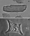
Fig. 1
Fig. 1
a The adult cardiomyocyte is approximately 25 μm in diameter and about 100 μm in length. It is composed of bundles of myofibrils that contain myofilaments. Myofibrils have distinct, repeating microanatomical units termed sarcomeres, which are the basic contractile elements that make up a developed cardiomyocyte. This conformation gives the adult cardiomyocyte its typical ‘rod-shape’ (‘cigar-shape’). b Unlike adult cardiomyocytes, which are highly organized and quite similar in morphology to one another, neonatal cardiomyocytes are in the process of developing their contractile machinery and therefore display a large variety of shapes. The neonatal cardiomyocyte is generally unable to retract its cell boundary during contraction, and noticeable changes occur only within the cell perimeter. For these reasons, it is difficult to perform contractile measurements on this cell type in a manner similar to that of the adult cardiomyocyte, in which changes in the cell boundary can be quantified during contraction
Fig. 2
Fig. 2
In the IonOptix® Myocyte Contractility Recording System, the SoftEdge® acquisition module collects cell length at rates up to 250 Hz. The simple user interface enables the precise placement of thresholds for marking cell edges. The output of calibrated length allows real-time cell length collection. Similarly, the sarcormere acquisition module collects real-time sarcomere length at 250 Hz. Line intensity information within a simple region of interest is averaged to generate a well-resolved striation pattern. A fast Fourier transform calculation immediately outputs sarcomere spacing. Along with the fast acquisition MyoCam-S® digital camera, the systems offer precise, real-time calcium and contractility measurements. Reproduced with permission from IonOptix®
Fig. 3
Fig. 3
Scanning electron micrograph of a PDMS elastomer micropillar array. In this case, the cross-sections of the pillars were close to square at the base and rounded toward the apex. Additionally, the cross-sectional areas normally increase slightly with height. (Reprinted from Kajzar et al. (2008) with permission from Elsevier)
Fig. 4
Fig. 4
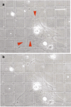
a Fibroblast before relaxation. Phase-contrast image of a rat cardiac fibroblast plated on a large grid pattern. As it contrasts, the cell creates distortions (arrowheads) by applying force to the elastomer (Young’s modulus = 18 kPa). b Fibroblast after relaxation. The same cell as in (a) 10 min after butanedione monoxime-induced relaxation. We observe the recovery of the regular grid pattern. Grid pitch = 30 mm. (Reprinted from Balaban et al. (2001) with permission from Macmillan)
Fig. 5
Fig. 5
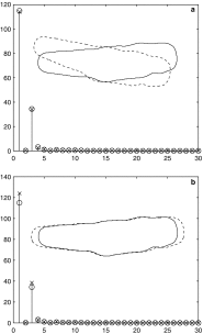
a Two identical cell shapes, one of which has been translated and rotated with respect to the other. First 30 Fourier descriptors of the shapes superimposed for the case of translation, rotation, and starting point invariance. b Two cell shapes, one of which is larger than the other, mimicking the ones from a relaxed and contracted cardiomyocyte, respectively. First 30 Fourier descriptors of the shapes superimposed. We observe that the Fourier descriptors are able to capture this change in shape size making the Fourier descriptors variant to scale but invariant to translation, rotation, and starting point. The ‘contraction’ of the shape is 8.15 %, as measured by the Euclidean distance of the Fourier descriptors. (Reprinted from Bazan et al. (2009) with permission from Hindawi)
Fig. 6
Fig. 6
Schematic of an atomic force microscope setup. The deflection of a micro-fabricated cantilever with a sharp tip (probe) at its end is quantified according to Hooke’s law by reflecting a laser beam off the backside of the cantilever into an array of photodiodes while it is scanning over the surface of the specimen. This figure is licensed by Grzegorz Wielgoszewski under the Creative Commons Attribution-Share Alike 3.0 Unported license
Fig. 7
Fig. 7
Schematic of a scanning ion-conductance microscope setup. An electrically-charged glass micropipette (or nanopipette) probe filled with electrolyte is lowered toward the surface of the specimen that is submerged in an oppositely-charged bath of electrolytes. As the micropipette gets closer to the specimen, the ion conductance decreases proportionally to the decrease in the distance between the two elements. These variations in the ion current are amplified and the signal is used to keep a constant distance between the micropipette and the specimen. The surface of the specimen can then be recorded. This figure was released into the public domain by Paul Venter
Fig. 8
Fig. 8
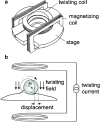
a Schematic of a microscope stage with twisting coils and magnetization coils. b A homogeneous magnetic twisting field provokes the bead to rotate and displace. The direction of the bead’s magnetic moment is denoted by M. (Reprinted from Fabry et al. (2001) with permission from The American Physiological Society)
Fig. 9
Fig. 9
Two micropipettes in a chamber. A pneumatic micromanipulator controls the movement of a micropipette along three orthogonal axes. a A micropipette exerts a suction pressure P to aspirate a spherical cell. b An attached cell being aspirated into a pipette. c A closely-fitting cell or bead moving freely in a pipette like a piston in a cylinder. When static, the suction pressure times the cross-sectional area of the pipette equals the attachment force F. (Reprinted from Hochmuth (2000) with permission from Elsevier)
Fig. 10
Fig. 10
Novel cell attachment clamp. A cell was loaded and the carbon fiber was lowered onto the cell. a A myocyte attached between the cantilevers. b Scanning electron microscopy image of the carbon fiber. (Reprinted from Garcia-Webb et al. (2007) with permission from The American Physiological Society)

References

    1. Addae-Mensah K, Wikswo J. Measurement techniques for cellular biomechanics in vitro. Exp Biol Med. 2008;233(7):792–809. doi: 10.3181/0710-MR-278. - DOI - PMC - PubMed
    1. Azeloglu E, Costa KD. Cross-bridge cycling gives rise to spatiotemporal heterogeneity of dynamic subcellular mechanics in cardiac myocytes probed with atomic force microscopy. Am J Physiol Heart Circ Physiol. 2010;298(3):H853–H860. doi: 10.1152/ajpheart.00427.2009. - DOI - PubMed
    1. Balaban N, Schwarz U, Riveline D, Goichberg P, Tzur G, Sabanay I, Mahalu D, Safran S, Bershadsky A, Addadi L, Geiger B. Force and focal adhesion assembly: a close relationship studied using elastic micropatterned substrates. Nat Cell Biol. 2001;3(5):466–472. doi: 10.1038/35074532. - DOI - PubMed
    1. Banyasz T, Lozinskiy H, Payne C, Edelmann S, Norton B, Chen B, Chen-Izu Y, Izu L, Balke C. Transformation of adult rat cardiac myocytes in primary culture. Exp Physiol. 2007;93(3):370–382. doi: 10.1113/expphysiol.2007.040659. - DOI - PubMed
    1. Bao G, Suresh S. Cell and molecular mechanics of biological materials. Nat Mater. 2003;2:715–725. doi: 10.1038/nmat1001. - DOI - PubMed

LinkOut - more resources