

Seeing the light with BLUF proteins
- PMID: 28510088
- PMCID: PMC5425820
- DOI: 10.1007/s12551-017-0258-6
Seeing the light with BLUF proteins
Abstract
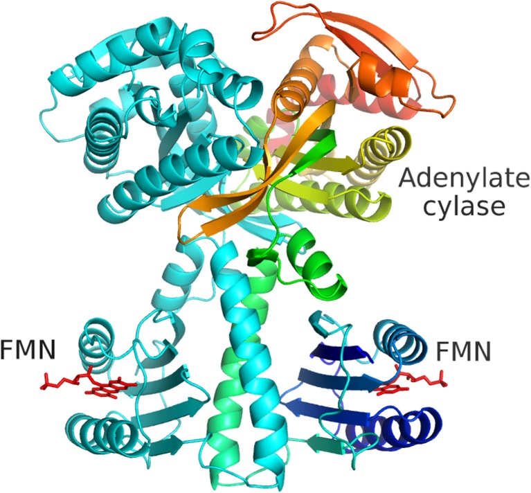
First described about 15 years ago, BLUF (Blue Light Using Flavin) domains are light-triggered switches that control enzyme activity or gene expression in response to blue light, remaining activated for seconds or even minutes after stimulation. The conserved, ferredoxin-like fold holds a flavin chromophore that captures the light and somehow triggers downstream events. BLUF proteins are found in both prokaryotes and eukaryotes and have a variety of architectures and oligomeric forms, but the BLUF domain itself seems to have a well-preserved structure and mechanism that have been the focus of intense study for a number of years. Crystallographic and NMR structures of BLUF domains have been solved, but the conflicting models have led to considerable debate about the atomic details of photo-activation. Advanced spectroscopic and computational methods have been used to analyse the early events after photon absorption, but these too have led to widely differing conclusions. New structural models are improving our understanding of the details of the mechanism and may lead to novel tailor-made tools for optogenetics.
Keywords: Allostery; Flavin; Optogenetics; Photo-activation.
Conflict of interest statement
Conflict of interest
Sam-Yong Park declares that he has no conflicts of interest.
Jeremy R. H. Tame declares that he has no conflicts of interest.
Ethical approval
This article does not contain any studies with human participants or animals performed by any of the authors.
Figures
Similar articles
-
Structure and insight into blue light-induced changes in the BlrP1 BLUF domain.Biochemistry. 2009 Mar 31;48(12):2620-9. doi: 10.1021/bi802237r. Biochemistry. 2009. PMID: 19191473
-
DFT/MM description of flavin IR spectra in BLUF domains.J Phys Chem B. 2011 Sep 29;115(38):11239-53. doi: 10.1021/jp2043637. Epub 2011 Sep 2. J Phys Chem B. 2011. PMID: 21888341
-
Light-induced structural changes in a putative blue-light receptor with a novel FAD binding fold sensor of blue-light using FAD (BLUF); Slr1694 of synechocystis sp. PCC6803.Biochemistry. 2004 May 11;43(18):5304-13. doi: 10.1021/bi049836v. Biochemistry. 2004. PMID: 15122896
-
Light detection and signal transduction in the BLUF photoreceptors.Plant Cell Physiol. 2013 Feb;54(2):171-9. doi: 10.1093/pcp/pcs173. Epub 2012 Dec 14. Plant Cell Physiol. 2013. PMID: 23243105 Review.
-
Time-resolved diffusion reveals photoreactions of BLUF proteins with similar functional domains.Photochem Photobiol Sci. 2022 Apr;21(4):493-507. doi: 10.1007/s43630-022-00214-2. Epub 2022 Apr 7. Photochem Photobiol Sci. 2022. PMID: 35391638 Review.
Cited by
-
Photoactivated Adenylyl Cyclases: Fundamental Properties and Applications.Adv Exp Med Biol. 2021;1293:129-139. doi: 10.1007/978-981-15-8763-4_7. Adv Exp Med Biol. 2021. PMID: 33398810 Review.
-
Optobiochemistry: Genetically Encoded Control of Protein Activity by Light.Annu Rev Biochem. 2021 Jun 20;90:475-501. doi: 10.1146/annurev-biochem-072420-112431. Epub 2021 Mar 29. Annu Rev Biochem. 2021. PMID: 33781076 Free PMC article. Review.
-
Current Trends of Bacterial and Fungal Optoproteins for Novel Optical Applications.Int J Mol Sci. 2023 Sep 29;24(19):14741. doi: 10.3390/ijms241914741. Int J Mol Sci. 2023. PMID: 37834188 Free PMC article. Review.
-
Toward Multiplexed Optogenetic Circuits.Front Bioeng Biotechnol. 2022 Jan 5;9:804563. doi: 10.3389/fbioe.2021.804563. eCollection 2021. Front Bioeng Biotechnol. 2022. PMID: 35071213 Free PMC article. Review.
-
Biophysical Reviews-A call to young biophysicists.Biophys Rev. 2021 May 28;13(3):289-294. doi: 10.1007/s12551-021-00810-z. eCollection 2021 Jun. Biophys Rev. 2021. PMID: 34178166 Free PMC article.
References
-
- Barends TRM, Hartmann E, Griese JJ et al (2009) Structure and mechanism of a bacterial light-regulated cyclic nucleotide phosphodiesterase. Nature 459:1015–1018 - PubMed
-
- Bonetti C, Stierl M, Mathes T et al (2009) The role of key amino acids in the photoactivation pathway of the Synechocystis Slr 1694 BLUF domain. Biochemistry 48:11458–11469 - PubMed
Publication types
LinkOut - more resources
Full Text Sources
Other Literature Sources
