Review
. 2015 Dec;7(4):353-368.
doi: 10.1007/s12551-015-0176-4. Epub 2015 Aug 23.

Biophysical chemistry of the ageing eye lens

Affiliations
Review

Biophysical chemistry of the ageing eye lens

Nicholas J Ray. Biophys Rev. 2015 Dec.

Abstract

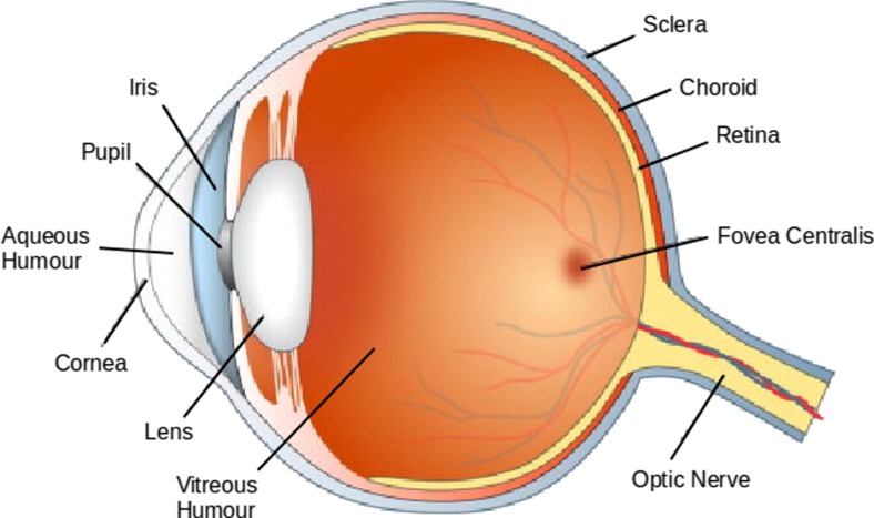
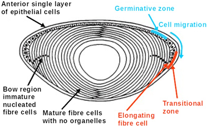
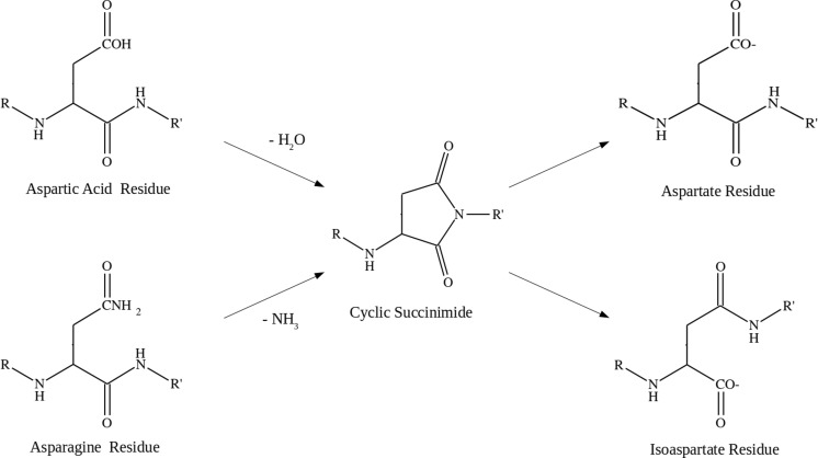
This review examines both recent and historical literature related to the biophysical chemistry of the proteins in the ageing eye, with a particular focus on cataract development. The lens is a vital component of the eye, acting as an optical focusing device to form clear images on the retina. The lens maintains the necessary high transparency and refractive index by expressing crystallin proteins in high concentration and eliminating all large cellular structures that may cause light scattering. This has the consequence of eliminating lens fibre cell metabolism and results in mature lens fibre cells having no mechanism for protein expression and a complete absence of protein recycling or turnover. As a result, the crystallins are some of the oldest proteins in the human body. Lack of protein repair or recycling means the lens tends to accumulate damage with age in the form of protein post-translational modifications. The crystallins can be subject to a wide range of age-related changes, including isomerisation, deamidation and racemisation. Many of these modification are highly correlated with cataract formation and represent a biochemical mechanism for age-related blindness.

Keywords: Ageing; Cataract; Crystallin; Eye lens; Post-tanslational modification.


Figures

Fig. 1
Fig. 1
Diagram of the human eye with major components labelled (Frost et al. 2014)
Fig. 2
Fig. 2
Cross-section of the human eye lens showing layers of lens fibre cells, the epithelial cell layer and the lens nucleus. Fibre cells differentiate form the epithelial layer and migrate to the lens equator. Mature fibre cells elongate and undergo denucleation (Moreau and King 2012)
Fig. 3
Fig. 3
Protein folding, unfolding and off-folding (aggregation) pathways (Ecroyd and Carver 2009)
Fig. 4
Fig. 4
sHSP interaction with unfolding and amorphous aggregation (Ecroyd and Carver 2009)
Fig. 5
Fig. 5
Chromatogram of lens fibre cells cytoplasm from bovine eye lens separated with gel filtration chromatography. Significant protein groups are marked. α- and β-crystallins separate into high and low mass elution peaks corresponding to different tertiary structures and γS-crystallin elutes separately to the other γ-crystallins
Fig. 6
Fig. 6
General structure of small heat shock proteins. Sequence of αB-crystallin (red) overlaid onto two related proteins, wheat sHSP 16.9 (green) and bacterial sHSP 16.5 (blue) (Ghosh and Clark 2005)
Fig. 7
Fig. 7
The structures of selected crystallin proteins. a NMR derived structure of human γS-crystallin two domain monomer (PDB 2M3T). b Crystal structure of bovine βB2-crystallin (PDB 1BLB) dimer showing domain sharing. c Crystal structure of human βB3-crystallin trimer (PDB 3QK3) with lattice structure
Fig. 8
Fig. 8
Mechanism for spontaneous cleavage of peptide chains at serine residues. The peptide chain undergoes rearrangement via N, O-acyl shift involving the HO-side-chain. Hydrolysis of the resulting ester results in truncation (Lyons et al. 2011)
Fig. 9
Fig. 9
Mechanism of deamidation in asparagine and aspartic acid residues via cyclic succinimide intermediate. Subsequent ring opening can produce both aspartic acid (α-amino acid) and isoaspartic acid (β-amino acid) products (Reissner and Aswad 2003)

References

    1. Aarts HJ, Lubsen NH, Schoenmakers JG. Crystallin gene expression during rat lens development. Eur J Biochem. 1989;183:31–36. doi: 10.1111/j.1432-1033.1989.tb14892.x. - DOI - PubMed
    1. Alizadeh A, Clark JI, Seeberger T, Hess J, Blankenship T, Spicer A, FitzGerald PG. Targeted genomic deletion of the lens-specific intermediate filament protein CP49. Invest Ophthalmol Vis Sci. 2002;43:3722–3727. - PubMed
    1. Allen D, Vasavada A. Cataract and surgery for cataract. Br Med J. 2006;333:128–132. doi: 10.1136/bmj.333.7559.128. - DOI - PMC - PubMed
    1. Augusteyn RC. On the growth and internal structure of the human lens. Exp Eye Res. 2010;90:643–654. doi: 10.1016/j.exer.2010.01.013. - DOI - PMC - PubMed
    1. Banh A, Bantseeva V, Choh V, Moran KL, Sivak JG. The lens of the eye as a focusing device and its response to stress. Prog Retin Eye Res. 2006;25:189–206. doi: 10.1016/j.preteyeres.2005.10.001. - DOI - PubMed