Review
. 2009 Mar;1(1):43.
doi: 10.1007/s12551-008-0007-y. Epub 2009 Jan 22.

Intercalated discs: multiple proteins perform multiple functions in non-failing and failing human hearts

Affiliations
Review

Intercalated discs: multiple proteins perform multiple functions in non-failing and failing human hearts

Colleen B Estigoy et al. Biophys Rev. 2009 Mar.

Abstract

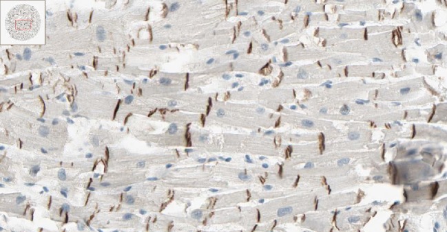
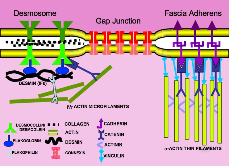
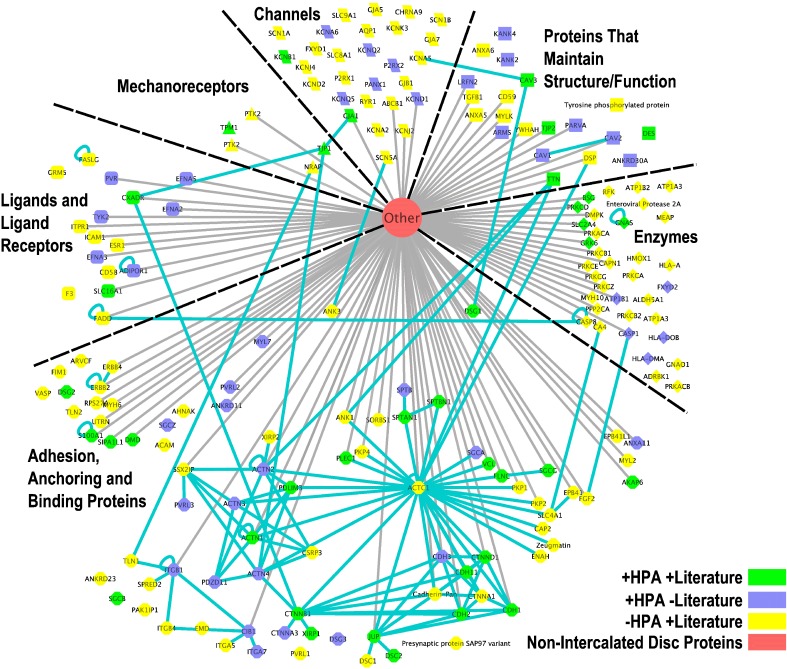
The intercalated disc (ICD) occupies a central position in the transmission of force, electrical continuity and chemical communication between cardiomyocytes. Changes in its structure and composition are strongly implicated in heart failure. ICD functions include: maintenance of electrical continuity across the ICD; physical links between membranes and the cytoskeleton; intercellular adhesion; maintenance of ICD structure and function; and growth. About 200 known proteins are associated with ICDs, 40% of which change in disease. We systemically reviewed cardiac immunohistochemical data on the Human Protein Atlas (HPA) web site, ExPASy protein binding data and published papers on ICDs. We identified 43 proteins not previously reported, and confirmed 37 proteins that have previously been described. In addition, 102 proteins not present on the HPA web site but were described in ICDs in the literature. We group these into clusters that demonstrate functionally interactive groups of proteins demonstrating that ICDs play a key role in cardiomyocyte function.

Keywords: Cardiac intercalated disc; Changes in disease; Functional protein groups; Human heart; Immunohistochemistry.


Figures

Fig. 1
Fig. 1
Immunohistochemistry of a human left ventricle stained with affinity-purified rabbit polyclonal antibody directed against a synthetic peptide that uniquely interacts with human desmoglein. Desmoglein was labelled using an HRP-labelled (brown) antibody. The nuclei are stained blue. The insert image in the top left corner shows a transverse section of a 1-mm diameter cylinder or core of paraffin-embedded left ventricle at high magnification. This image can be viewed on the HPA web site located at: (http://www.proteinatlas.org/normal_unit.php?antibody_id=4896&mainannotation_id=633425)
Fig. 2
Fig. 2
A diagrammatic representation of three structural zones of the intercalated disc. Colours above and below the ICD are shaded darker and lighter to indicate adjacent myoplasms. Note that the fascia adherens mainly anchors myofibrillar proteins, whilst the desmosome is mainly linked to the cytoskeleton (intermediate filaments and microfilaments). Five connexon channels are shown in the gap junction where each connexon composed of six subunits, usually comprising connexin 40 and 43. These proteins are summarised in recent reviews (Kostin et al ; Zuppinger et al 2000)
Fig. 3
Fig. 3
Visualisation of the interaction network of the intercalated disc proteins. This figure was produced by Cytoscape (Shannon et al. 2003). The nodes are coloured according to the level of evidence for the presence of each protein in the intercalated disc. All nodes are grouped according to the functional categories listed in Electronic supplementary material Table 1. All proteins in the same functional category have the same node shape. An interaction between a pair of intercalated disc proteins is drawn as a solid blue line, whilst an interaction between an ICD protein and a non-ICD protein (represented by the red “Other” node in the figure) is drawn as a grey line. Proteins that interact with themselves are indicated by a loop. Proteins that have no connecting line have simply been identified in the ICD but have no known interactions with other proteins

References

    1. Basso C, Czarnowska E, Barbera MD, et al. Ultrastructural evidence of intercalated disc remodelling in arrhythmogenic right ventricular cardiomyopathy: an electron microscopy investigation on endomyocardial biopsies. Eur Heart J. 2006;27:1847–1854. doi: 10.1093/eurheartj/ehl095. - DOI - PubMed
    1. Bennett PM, Maggs AM, Baines AJ, Pinder JC. The transitional junction: a new functional subcellular domain at the intercalated disc. Mol Biol Cell. 2006;17:2091–2100. doi: 10.1091/mbc.E05-12-1109. - DOI - PMC - PubMed
    1. Bjorling E, Lindskog C, Oksvold P, et al. A web-based tool for in silico biomarker discovery based on tissue-specific protein profiles in normal and cancer tissues. Mol Cell Proteomics. 2008;7:825–844. doi: 10.1007/978-1-59745-117-8. - DOI - PubMed
    1. Bourne GH. Enzymes of the intercalated disks of heart muscle. Nature. 1952;172:588–589. doi: 10.1038/172588a0. - DOI - PubMed
    1. dos Remedios CG, Chhabra D, Kekic M, Dedova IV, Tsubakihara M, Berry DA, Nosworthy NJ. Actin and actin binding proteins: regulation of cytoskeletal microfilaments. Physiol Rev. 2003;83:433–473. - PubMed

LinkOut - more resources