. 2017 Sep;17(9):2458-2467.
doi: 10.1111/ajt.14359. Epub 2017 Jun 27.

Proteoforms in Peripheral Blood Mononuclear Cells as Novel Rejection Biomarkers in Liver Transplant Recipients

Affiliations

Proteoforms in Peripheral Blood Mononuclear Cells as Novel Rejection Biomarkers in Liver Transplant Recipients

T K Toby et al. Am J Transplant. 2017 Sep.

Abstract

Biomarker profiles of acute rejection in liver transplant recipients could enhance the diagnosis and management of recipients. Our aim was to identify diagnostic proteoform signatures of acute rejection in circulating immune cells, using an emergent "top-down" proteomics methodology. We prepared differentially processed and cryopreserved cell lysates from 26 nonviral liver transplant recipients by molecular weight-based fractionation and analyzed them by mass spectrometry of whole proteins in three steps: (i) Nanocapillary liquid chromatography coupled with high-resolution tandem mass spectrometry; (ii) database searching to identify and characterize intact proteoforms; (iii) data processing through a hierarchical linear model matching the study design to quantify proteoform fold changes in patients with rejection versus normal liver function versus acute dysfunction without rejection. Differentially expressed proteoforms were seen in patients with rejection versus normal and nonspecific controls, most evidently in the cell preparations stored in traditional serum-rich media. Mapping analysis of these proteins back to genes through gene ontology and pathway analysis tools revealed multiple signaling pathways, including inflammation mediated by cytokines and chemokines. Larger studies are needed to validate these novel rejection signatures and test their predictive value for use in clinical management.

Keywords: biomarker; clinical research/practice; immunobiology; liver allograft function/dysfunction; liver transplantation/hepatology; monitoring: immune; proteomics; rejection: acute; translational research/science.


Conflict of interest statement

Disclosure

The authors of this manuscript have conflicts of interest to disclose as described by the American Journal of Transplantation. NLK declares an affiliation with Thermo-Fisher Scientific. JL and MA have affiliations with Transplant Genomics Incorporated. The other authors have no conflicts of interest to disclose.

Figures

Figure 1
Figure 1. Schematic workflow for discovery-mode, translational, top-down proteomics applied to liver transplant patient groups (acute rejection [AR]; transplant excellent [TX]; acute dysfunction, no rejection [ADNR]) from blood collection to informatics analysis
Label-free top-down quantitation in translational research is a complex methodology with many aspects. First, patients were stratified into three groups by independent pathologist review to confirm phenotypic assignments. TX patients showed no signs of allograft dysfunction, AR patients demonstrated rejection activity index scores ≥3, and ADNR patients were included as a nonspecific liver dysfunction control. In all three phenotypes, PBMCs were collected from eight to 10 patients per state. Proteins were resolved by size using gel-eluted liquid fraction entrapment electrophoresis, and the resulting fractions were analyzed by LC-MS in technical quadruplicate and forwarded for ANOVA analysis. LC, liquid chromatography; MS, mass spectrometry; PBMC, peripheral blood mononuclear cell. [Color figure can be viewed at wileyonlinelibrary.com]
Figure 2
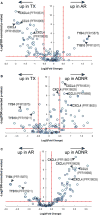
Figure 2. Label-free, top-down, quantitative analysis describing differentially expressed proteoforms for all three comparisons of the liver transplantation patient groups in this study (AR, TX, and ADNR)
For all proteoforms detected in the majority of data files across the data set (open circles), ANOVA was used to assign variation in signal intensity to phenotype-specific effects after accounting for patient-to-patient and technical variation. The x-axis represents the effect size as measured by fold-change (log2 transformed) between patient groups. The y-axis (FDR-corrected p-value) is a measure of the statistical confidence that signal variation is associated with phenotype. The dashed lines represent our arbitrary thresholds for delineating significant hits: The horizontal dashed line corresponds to a 5% FDR, and vertical dashed lines represent effect sizes 1.4-fold above and below no change. Arrowheads denote certain proteoforms of interest that are discussed in the text. ADNR, acute dysfunction, no rejection; AR, acute rejection; FDR, false discovery rate; PFR, proteoform record number; TX, transplant excellent; CCL, C-C motif chemokine ligand; TYB, thymosin β. [Color figure can be viewed at wileyonlinelibrary.com]
Figure 3
Figure 3. Comprehensive analysis of the posttranslational processing events characterized by top-down proteomics in the differentially expressed proteoforms filtered out of the AR versus TX quantitative comparison results
The entire list of differentially expressed proteoforms generated from the transplant excellent versus acute rejection comparison was manually annotated for chemical and sequence modifications that caused divergence from the canonical sequence associated with the protein accession number. The histogram (top panel) depicts the number of specific posttranslational processing events sorted into bins sorted by their prevalence in the data set. The identities of the identified posttranslational processing events are depicted in the table (bottom panel), sorted by event number on the histogram x-axis. The UniProt Knowledgebase (http://www.uniprot.org/) was the source for curating all annotated modifications described.
Figure 4
Figure 4. Proteoform-resolved analysis of PF4/CXCL4 differentially expressed proteoforms characterized by top-down proteomics and an in silico comparison to tryptic peptide-based approaches
(A) The full-length canonical sequence of PF4/CXCL4 (accession no. P02776). Blue flags depict the cleavage sites of the three differentially abundant PF4/CXCL4 proteoforms and are labeled with their identity. The sequence underlined in green is the signal peptide, and red arrows delineate trypsin cleavage sites generated in silico. Notably, tryptic peptides do not span the region of sequence variability describing the three proteoforms of interest in this study. (B) Box-and-whisker plot comparison of the 8140-Da PF4/CXCL4 proteoform intensities across all patients and injections, which were found to be significantly decreased in AR patients. (C) Box-and-whisker plots made from aggregating all PF4/CXCL4 proteoform intensities per patient group to emulate a quantitative comparison using intensities of tryptic peptides, which cannot distinguish the proteoforms. Notably, the effect size is lost to noise in this in silico experiment, and the analysis would return a false negative by bottom-up proteomics. For the box-and-whisker plots, data points represent the normalized intensities of the proteoform of interest yielded from every technical replicate (data file) per patient in which the proteoform was detected (TX: n = 8 patients, 31 data files; ADNR n = 9 patients, 31 data files; AR: n = 9 patients, 33 data files). ADNR, acute dysfunction, no rejection; AR, acute rejection; PF4, platelet factor 4; PFR, proteoform record number; TX, transplant excellent. [Color figure can be viewed at wileyonlinelibrary.com]

References

    1. Charlton M, Seaberg E. Impact of immunosuppression and acute rejection on recurrence of hepatitis C: Results of the national institute of diabetes and digestive and kidney diseases liver transplantation database. Liver Transpl Surg. 1999;5(4 Suppl 1):S107–S114. - PubMed
    1. Levitsky J, Goldberg D, Smith AR, et al. Acute rejection increases risk of graft failure and death in recent liver transplant recipients. Clin Gastroenterol Hepatol. 2016;15:584–593. - PMC - PubMed
    1. Shaked A, Chang BL, Barnes MR, et al. An ectopically expressed serum miRNA signature is prognostic, diagnostic, and biologically related to liver allograft rejection. Hepatology. 2017;65:269–280. - PubMed
    1. Massoud O, Heimbach J, Viker K, et al. Noninvasive diagnosis of acute cellular rejection in liver transplant recipients: A proteomic signature validated by enzyme-linked immunosorbent assay. Liver Transpl. 2011;17:723–732. - PMC - PubMed
    1. Moya-Quiles MR, Alvarez R, Miras M, et al. Impact of recipient HLA-C in liver transplant: A protective effect of HLA-Cw*07 on acute rejection. Hum Immunol. 2007;68:51–58. - PubMed