. 2014 Dec;55(1):28.
doi: 10.1186/1999-3110-55-28. Epub 2014 Mar 1.

From leaf and branch into a flower: Magnolia tells the story

Affiliations

From leaf and branch into a flower: Magnolia tells the story

Wen-Zhe Liu et al. Bot Stud. 2014 Dec.

Abstract

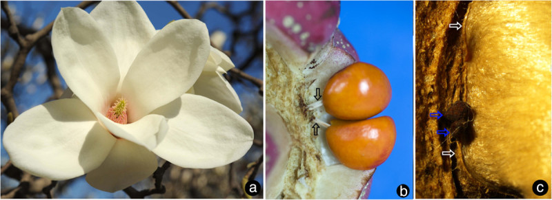
Background: In the classical doctrines, Magnolia was frequently considered the archetype among flowering plants, and its conduplicate carpel with marginal placentation was assumed to be derived from a leaf-like organ bearing ovules along its margins. Although the robustness of this concept has been seriously questioned by advances in botanical research, especially the emergence of Magnolia deeper in the angiosperm tree of life in molecular systematics, it remains the most-taught interpretation for the origin of carpels.

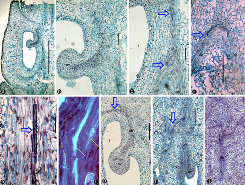
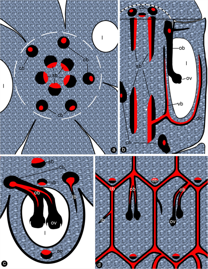
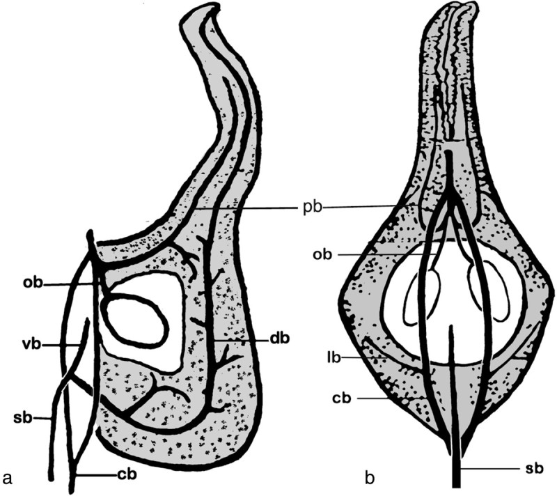
Results: To test the validity of this classical doctrine, we performed comparative anatomical analyses of the vascular bundles in the flowers of Magnolia using fine (8-μm) paraffin -sections. We document the presence of two independent vascular systems in the carpels: the collateral bundles of the dorsal and ventral veins arising from the stelar bundle, and the amphicribral ovular bundles arising from the cortical bundles. This observation in conjunction with data from other fields concurrently suggests that the ovary wall is equivalent to a foliar organ whereas the placenta represents an ovule-bearing shoot.

Conclusions: Our observation on the former model plant, Magnolia, nullifies the classical doctrine of carpel evolution and supports the Unifying Theory. This conclusion prompts a reconsideration of the concept of angiosperm flower evolution.

Keywords: Angiosperm; Carpel; Comparative anatomy; Flower; Magnolia; Origin.


Figures

Figure 1
Figure 1
Cross sections of the gynoecium in Magnolia denudata flower. cb: cortical bundle, db: dorsal bundle, ep: epidermis, l:ovarian locule, lb: lateral bundle, ob: ovular bundle, ov: ovule, sb: stelar bundle, vb: ventral bundle. Adaxial side is to the bottom. Specimen number NWU00032054-4403, deposited at Northwest University, China. a. Cross section of a flower, showing several carpels around the floral axis. Bar = 1 mm. b. Detailed view of the rectangle in Figure. 2a, showing cortical bundles giving rise to two ovular bundles as well as a lateral bundle. Bar = 0.2 mm. c. Detailed view of the rectangle in Figure 1b, showing two ovular bundles and a lateral bundle. Bar = 0.2 mm. d. Two ovules in the same ovary arising from the same ovular bundle (arrow). Bar = 0.2 mm. e. Floral axis with stelar bundles, cortical bundles, and their relationship with carpel boundaries and ovular bundles. Bar = 0.2 mm. f. Collateral stelar bundle with adaxial xylem (arrow). Bar = 50 μm. g. Amphicribral cortical bundle with close-to-center xylem (arrow). Bar = 50 μm.h. Collateral dorsal bundle with adaxial xylem (arrow). Bar = 50 μm. i. Three collateral stelar bundles with adaxial xylem (white arrows) and abaxial phloem (yellow arrows). Bar = 50 μm. j. Amphicribral ovular bundle with phloem (P) (yellow arrows) surrounding the xylem (X) in the center (circle). Bar = 50 μm. k. Amphicribral cortical bundle with phloem (yellow arrows) surrounding the xylem in the center (white). Bar = 0.1 mm.
Figure 2
Figure 2
Longitudinal sections of Magnolia denudata flower. For abbreviations, see caption for Figure 1. Specimen number NWU00032054-4405, deposited at Northwest University, China. a. Radial section showing a carpel and its relationship to the floral axis to the right. Bar = 1 mm. b. Detailed view of the ovule in Figure 2a. Note the relationship between the ovule, descending ovular bundle, and ventral bundle. Bar = 0.1 mm. c. Detailed view of the dorsal bundle in Figure 1a. Note the relationship among the ascending ventral bundle (vb, blue arrows), descending dorsal bundle (db, blue arrows), and ascending stelar bundle (sb). Bar = 0.1 mm. d. Dorsal (db, blue arrows) and stelar bundles (sb) in another carpel. Bar = 0.2 mm. e. Vessel element (blue arrow) in an amphicribral cortical bundle, with central xylem. Bar = 50 μm. f. Xylem (tracheids, white arrow) sandwiched between phloem (sieve elements, yellow arrows) in the amphicribral ovular bundle. Bar = 0.1 mm. g. An ovular bundle (ob) supplying the ovule and ascending along the carpel margin (blue arrow). Bar = 0.2 mm. h. Tangential section showing anastomosing cortical bundles (cb) connected to ovular bundles (ob). Note the branching (blue arrow) of the cortical bundles, and their relationship to the ovular bundles. Bar = 0.2 mm. i. Tangential section showing a dorsal bundle branching pinnately. Bar = 0.2 mm.
Figure 3
Figure 3
Flower and fruits of Magnolia denudata . a. A blooming flower. b. Red seeds hanging on the placenta after the ovary wall is shed. Note the connections (arrows) between the placenta and seeds. c. Two aborted ovules (blue arrows) independent of their ovary wall in the background. Note the border of the ovary wall (white arrow).
Figure 4
Figure 4
Sketches showing the vascular anatomy of Magnolia denudata. For abbreviations, see caption for Figure 1. Red color for xylem, and black for phloem. a. Cross section showing ovarian locules, carpels around the floral axis (white broken line), collateral stelar bundles in the center, and amphicribral cortical bundles in the cortex. b. Three dimensional diagram showing a carpel attached to the floral axis, collateral stelar and amphicribral cortical bundles, ventral and dorsal bundles derived from the stelar bundles. c. Cross section of a carpel showing the deployment of collateral stelar bundle, collateral ventral and dorsal bundles, amphicribral cortical and ovular bundles, and lateral bundle. d. Anastomosis of cortical bundles and their relationship with ovules and lateral bundles in carpels.
Figure 5
Figure 5
Diagrams of vascular patterns of carpels in Magnolia denudata (redrawn and modified from Canright, 1960). cb: cortical bundle, db: dorsal bundle, lb: lateral bundle, ov: ovule, ob: ovular bundle, pb: placenta bundle, sb: stelar bundle, vb: ventral bundle. a. The lateral view. b. The ventral view, look into the flower periphery.
Figure 6
Figure 6
Flower and its anatomy of Magnolia championii. cb: cortical bundle, l:ovarian locule, lb: lateral bundle, ob: ovular bundle, ov: ovule, vb: ventral bundle. a. Blooming flower. b. Longitudinal sections of a flower, showing relationship between ovarian locule, ovule, ovular bundle, and ventral bundle. c. Detailed view of Figure 6b, showing ovular and ventral bundles. d. The central xylem (white arrow) in ovular bundle and abaxial xylem (black arrow) in ventral bundle. e. Cross section of an amphicribral ovular bundle with central xylem (white arrow) in the placenta. f-g. Longitudinal sections of carpels in their early development showing carpel wall (CW) and placenta (arrows).

References

    1. Angenent GC, Franken J, Busscher M, Van Dijken A, Van Went JL, Dons H, Van Tunen AJ. A novel class of MADS box genes is involved in ovule development in Petunia. Plant Cell. 1995;7:1569–1582. doi: 10.1105/tpc.7.10.1569. - DOI - PMC - PubMed
    1. APG An update of the Angiosperm Phylogeny Group classification for the orders and families of flowering plants: APG II. Bot J Linn Soc. 2003;141:399–436. doi: 10.1046/j.1095-8339.2003.t01-1-00158.x. - DOI
    1. APG An update of the Angiosperm Phylogeny Group classification for the orders and families of flowering plants: APG III. Bot J Linn Soc. 2009;161:105–121. doi: 10.1111/j.1095-8339.2009.00996.x. - DOI
    1. Arber A. The natural philosophy of plant form. Cambridge: University Press; 1950.
    1. Bailey IW, Nast CG. The comparative morphology of the Winteraceae II. Carpels. J Arnold Arboretum. 1943;24:472–481.

LinkOut - more resources