. 2018 Jan;38(1):58-74.
doi: 10.1177/0271678X17705973. Epub 2017 May 17.

mTOR drives cerebral blood flow and memory deficits in LDLR-/- mice modeling atherosclerosis and vascular cognitive impairment

Affiliations

mTOR drives cerebral blood flow and memory deficits in LDLR-/- mice modeling atherosclerosis and vascular cognitive impairment

Jordan B Jahrling et al. J Cereb Blood Flow Metab. 2018 Jan.

Abstract

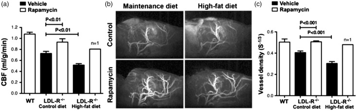
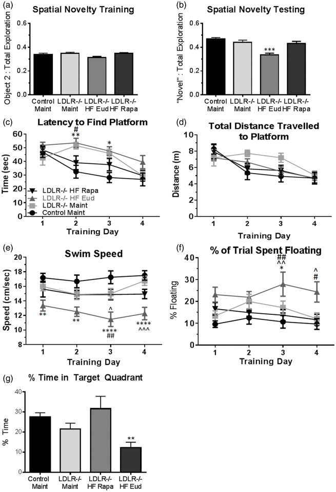
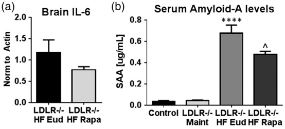
We recently showed that mTOR attenuation blocks progression and abrogates established cognitive deficits in Alzheimer's disease (AD) mouse models. These outcomes were associated with the restoration of cerebral blood flow (CBF) and brain vascular density (BVD) resulting from relief of mTOR inhibition of NO release. Recent reports suggested a role of mTOR in atherosclerosis. Because mTOR drives aging and vascular dysfunction is a universal feature of aging, we hypothesized that mTOR may contribute to brain vascular and cognitive dysfunction associated with atherosclerosis. We measured CBF, BVD, cognitive function, markers of inflammation, and parameters of cardiovascular disease in LDLR-/- mice fed maintenance or high-fat diet ± rapamycin. Cardiovascular pathologies were proportional to severity of brain vascular dysfunction. Aortic atheromas were reduced, CBF and BVD were restored, and cognitive dysfunction was attenuated potentially through reduction in systemic and brain inflammation following chronic mTOR attenuation. Our studies suggest that mTOR regulates vascular integrity and function and that mTOR attenuation may restore neurovascular function and cardiovascular health. Together with our previous studies in AD models, our data suggest mTOR-driven vascular damage may be a mechanism shared by age-associated neurological diseases. Therefore, mTOR attenuation may have promise for treatment of cognitive impairment in atherosclerosis.

Keywords: Atherosclerosis; cerebral blood flow; cognition; inflammation; vascular biology.


Figures

Figure 1.
Figure 1.
Chronic rapamycin treatment reduces mTOR activity in aortas and brain vasculature of LDLR−/− mice. (a) Blood levels of rapamycin in mice of Cohort 1 after 30 weeks of treatment. Blood levels of control-treated animals were below the detection limit (D.L.) of 0.5 ng/ml. (b) Significantly reduced phosphorylation of rpS6, a downstream target of mTOR, in aortas from rapamycin-treated animals (**, p = 0.004, Student’s unpaired t test); (c) Significantly reduced rpS6 phosphorylation in brain vasculature purified from rapamycin-treated animals of Cohort 4 following 20 weeks of treatment (***, F(3,12) = 12.06, p < 0.001, Tukey’s test on a significant effect of treatment, one-way ANOVA); (d) Rapamycin levels in liver in animals of Cohort 4. n = 8–10/group. Data are means ± SEM.
Figure 2.
Figure 2.
Chronic mTOR attenuation reduces body weight and fat mass gains in HFD-fed LDLR−/− mice. (a) Decreased body weight despite a significant increase in blood triglycerides without changes in glucose or cholesterol in rapamycin-treated HFD-fed LDLR−/− mice of Cohort 1; (b) Reduced body fat in rapamycin-treated HFD-fed LDLR−/− mice of Cohort 1 (*, p = 0.0465, Student’s unpaired t test); (c) Weight gain in Cohort 4 HFD- two-way ANOVA); (d) Reduced body fat in rapamycin-treated LDLR−/− mice of Cohort 4 (**, F(3, 42) = 38.54, p < 0.0001, Tukey’s test on a significant effect of treatment, one-way ANOVA); No differences in food consumption (e) or fasting glucose (f) levels were observed among experimental groups; (g) Significant increases in blood cholesterol following HFD feeding in Cohort 4 were unaffected by rapamycin treatment. n = 11–15/group (males) and n = 13–16/group (females) for (a–b) and n = 10/group (c–f). Data are means ± SEM.
Figure 3.
Figure 3.
Reduced atherosclerotic lesion size in the aortic arch following mTOR attenuation. Overall aortic arch lesion area (a) in HFD-fed LDLR−/− female (left panel), male (center panel) and both female and male (right panel) mice were significantly reduced following either 20 or 30 weeks of rapamycin treatment (*, two-way ANOVA F(1,34) = 11.69, p = 0.002) n = 6–8/group; (b) Reduced lesion area by rapamycin treatment in the aortic root of female mice after, but not prior to, the onset of lesion formation (left panel, quantitative analyses of anatomical data; right panel, representative images (oil red o (ORO), haemotoxylin and eosin (H&E)). Student’s unpaired t test, *, p = 0.026), n = 12/group (c) No difference in male but significantly reduced left ventricle mass in female LDLR −/− mice treated with rapamycin (*, two-way ANOVA F(1,28) = 10.30, p = 0.029), n = 8/group. Data are means ± SEM.
Figure 4.
Figure 4.
Restored cerebral blood flow and preserved brain vascular density by mTOR attenuation in LDLR−/− mice. (a) Profound CBF deficits in 12 month old LDLR−/− mice were restored to levels indistinguishable from those of WT mice as a result of chronic rapamycin treatment (F(2,7) = 21.22, p = 0.001, Tukey’s test, one-way ANOVA); (b–c) Reduced BVD (calculated as S = ΔR2/(ΔR2*)2/3) in control-treated but not in rapamycin-treated LDLR−/− mice (F(2,7) = 41.48, p = 0.0001, Tukey’s test, one-way ANOVA; (b) Representative MRI angiograms; (c) Quantitative analyses of angiogram data. n = 3–4. Data are means ± SEM. N.B. Data for the single surviving HFD-fed rapamycin-treated LDLR−/− animal are provided (a,c) but were not included in the ANOVA. Values for WT CBF (a) and BVD (c) are included as a reference and correspond to measures performed on separate groups of WT C57Bl/6 J mice of comparable age; these values were not included in the ANOVA.
Figure 5.
Figure 5.
Rapamycin treatment abolishes memory impairments in LDLR−/− mice. (a) No differences among experimental groups during training in the spatial novelty task; (b) Recognition memory in rapamycin-treated HFD-fed LDLR−/− mice is restored to levels indistinguishable from those of WT mice (F(3, 35) = 7.048, p = 0.0008 Tukey’s test, one-way ANOVA), n = 10–11/group; (c) Improved performance in rapamycin-treated HFD-fed mice, but not in in control-treated LDLR−/− mice fed either HFD or maintenance chow in days 2 and 3 of training in the MWM [(F(3, 35) = 6.336, p = 0.0015) and training day (F (3, 105) = 15.19, p < 0.0001, Tukey’s test, RM two-way ANOVA]. Although all groups swam comparable distances during training days, distances swam were artificially decreased in untreated HFD-fed LDLR−/− mice (d) due to a significant reduction in swimming speed (e) (F(3, 35) = 10.71, p < 0.0001, Tukey’s test, RM two-way ANOVA) as a result of increased floating (f) (F (3, 35) = 4.87, p = 0.006, Tukey’s test, RM two-way ANOVA). n = 10/group. Data are means ± SEM. (g) HFD-fed LDLR−/− mice exhibit significant cognitive impairment during the probe trial in which the platform is removed. Rapamycin treatment restored exploration in the appropriate quadrant to WT levels (**, one-way ANOVA, p = 0.013). *, HFD-fed LDLR−/− different from Control Maint; ^, HFD-fed LDLR−/− different from Maint-fed LDLR−/−; #, HFD-fed LDLR−/− different from rapa-fed LDLR−/− mice.
Figure 6.
Figure 6.
Chronic rapamycin treatment abrogates an increase in markers of systemic inflammation in LDLR−/− mice. (a) Reduced brain IL-6 levels in rapamycin-treated HFD-fed LDLR−/− mice as compared to control-treated HFD-fed LDLR−/− mice [p = 0.2566, Student’s unpaired t test with Welch’s correction. Variance was significantly different among groups (p = 0.001, F(4, 6) = 12.09], n = 5–7/group); (b) Increased serum levels of SAA in control HFD-fed LDLR −/− mice as compared to maintenance-fed LDLR−/− are attenuated by rapamycin treatment (****p < 0.0001, F(3, 15) = 49.79, Tukey’s test, one-way ANOVA and p = 0.028, Student’s unpaired t test). n = 6/group Data are means ± SEM.

References

    1. Morris JC. Early-stage and preclinical Alzheimer disease. Alzheimer Dis Assoc Disord 2005; 19: 163–165. - PubMed
    1. Breteler MM, Claus JJ, Grobbee DE, et al. Cardiovascular disease and distribution of cognitive function in elderly people: the Rotterdam Study. BMJ 1994; 308: 1604–1608. - PMC - PubMed
    1. Kuller LH, Shemanski L, Manolio T, et al. Relationship between ApoE, MRI findings, and cognitive function in the Cardiovascular Health Study. Stroke 1998; 29: 388–398. - PubMed
    1. Kivipelto M, Ngandu T, Fratiglioni L, et al. Obesity and vascular risk factors at midlife and the risk of dementia and Alzheimer disease. Arch Neurol 2005; 62: 1556–1560. - PubMed
    1. Zlokovic BV. Neurovascular pathways to neurodegeneration in Alzheimer's disease and other disorders. Nat Rev Neurosci 2011; 12: 723–738. - PMC - PubMed

Publication types

MeSH terms

LinkOut - more resources