. 2017 May 16;5(1):56.
doi: 10.1186/s40168-017-0273-7.

Tomato root microbiota and Phytophthora parasitica-associated disease

Affiliations

Tomato root microbiota and Phytophthora parasitica-associated disease

Marie Larousse et al. Microbiome. .

Abstract

Background: Interactions between pathogenic oomycetes and microbiota residing on the surface of the host plant root are unknown, despite being critical to inoculum constitution. The nature of these interactions was explored for the polyphagous and telluric species Phytophthora parasitica.

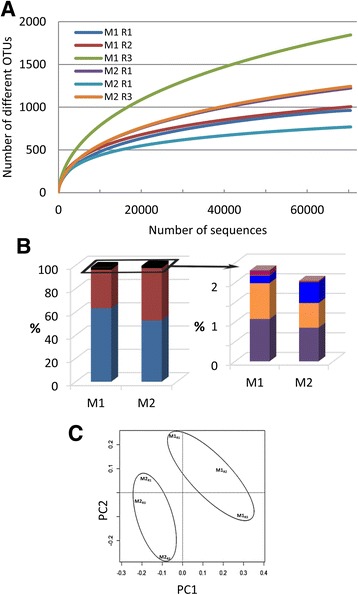
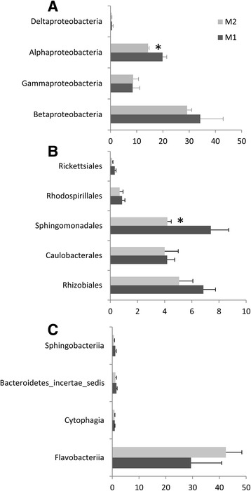
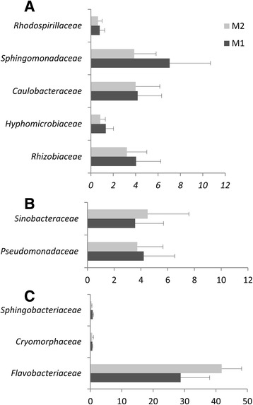
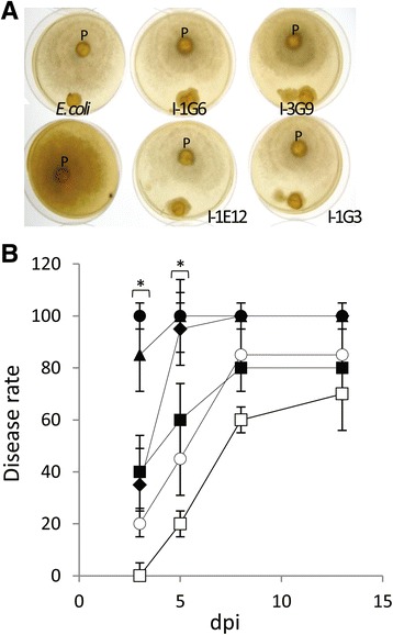
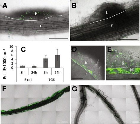
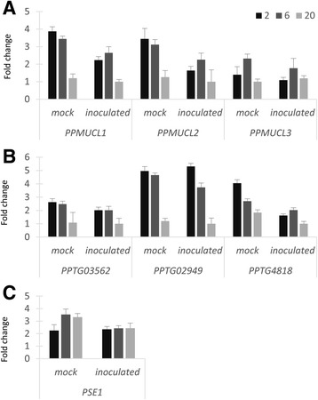
Results: Composition of the rhizospheric microbiota of Solanum lycopersicum was characterized using deep re-sequencing of 16S rRNA gene to analyze tomato roots either free of or partly covered with P. parasitica biofilm. Colonization of the host root surface by the oomycete was associated with a shift in microbial community involving a Bacteroidetes/Proteobacteria transition and Flavobacteriaceae as the most abundant family. Identification of members of the P. parasitica-associated microbiota interfering with biology and oomycete infection was carried out by screening for bacteria able to (i) grow on a P. parasitica extract-based medium (ii), exhibit in vitro probiotic or antibiotic activity towards the oomycete (iii), have an impact on the oomycete infection cycle in a tripartite interaction S. lycopersicum-P. parasitica-bacteria. One Pseudomonas phylotype was found to exacerbate disease symptoms in tomato plants. The lack of significant gene expression response of P. parasitica effectors to Pseudomonas suggested that the increase in plant susceptibility was not associated with an increase in virulence. Our results reveal that Pseudomonas spp. establishes commensal interactions with the oomycete. Bacteria preferentially colonize the surface of the biofilm rather than the roots, so that they can infect plant cells without any apparent infection of P. parasitica.

Conclusions: The presence of the pathogenic oomycete P. parasitica in the tomato rhizosphere leads to a shift in the rhizospheric microbiota composition. It contributes to the habitat extension of Pseudomonas species mediated through a physical association between the oomycete and the bacteria.

Keywords: Biofilm; Flavobacteriaceae; Host plant; Metagenomics 16S; Oomycete; Pseudomonadaceae.


Figures

Fig. 1
Fig. 1
a Rarefaction curves of observed OTUs richness for each of the three M1 and M2 replicates and using an OTU threshold of ≥97% identity. b Percentage of high-confidence OTUs grouped by phylum: Proteobacteria (blue) and Bacteroidetes (red) (left panel); Latescibacteria (purple), Actinobacteria (orange), Firmicutes (blue), Verrucomicrobia (dark pink) (right panel). c PCA ordination based on Hellinger distances
Fig. 2
Fig. 2
Relative abundance histograms (percentage ± SD) of Proteobacteria (a), Alphaproteobacteria orders (b), and Bacteroidetes classes (c) present in M1 or M2. Differences between M1 and M2 were significant for Alphaproteobacteria (p = 0.03) and Sphingomonadales (p = 0.05) in a Student’s t test (n = 3)
Fig. 3
Fig. 3
Relative abundance histograms (percentage + SD) of the ten most abundant families listed for M1 and M2 (threshold >0.5%) in the presence (light gray) and in the absence (dark gray) of P. parasitica biofilm. a Alphaproteobacteria. b Gammaproteobacteria. c Bacteroidetes
Fig. 4
Fig. 4
Bacterial isolate analyses. a Plate confrontation assay of the E. coli strain and the M2-isolates (I-1G6, I-3G9, I-1E12, and I-1G3) exhibiting antimicrobial activity against P. parasitica (P) performed on V8 extract agar medium. b Histogram of the disease rate measured at different days post zoospore inoculation (○) or during a tripartite interaction with E. coli (□),I-3G9 (●), I-1E12 (▲), I-1G6 (♦), or I-1G3 (■). Disease symptoms of individual plants were monitored at 3, 5, 8, and 13 days (dpi). I-3G9 as well as I-1E12 differed from E.coli and mock inoculation in disease index measured at 3 and 5 dpi (n = 5, Student’s t test, 0.004 < p < 0.035). Values are the means ± SD of two replicates
Fig. 5
Fig. 5
Location of I-1G6-GFP. Micrographs illustrating the preferential location of 1G6-GFP cells on biofilm (b) compared to root (r) at 3 hpi (a), 48 hpi (d, e), and 8 dpi (f). The colonization observed for I-1G6-GFP at 3 hpi (a) 8 dpi (f) can be compared to the lesser one observed for E. coli-GFP (b) and (g), respectively. c Histogram of the relative fluorescence intensity at 3 and 24 hpi for I-1G6-GFP and E.coli-GFP, measured as the ratio of the mean values at the surface of the biofilms and of roots. Bars: 200 μm in a, b, f, and g; 50 μm in d and e. The dotted lines delineate the interface between biofilm and root. In (e) dotted lines delineate an epidermal cell infected by I-1G6-GFP-and located beneath a biofilm
Fig. 6
Fig. 6
P. parasitica gene expression patterns in response to I-1G6-GFP. Roots of 2-week-old plants covered with a P. parasitica biofilm after 3 h of incubation with zoospores. They were then incubated in water or in the presence of I-1G6-GFP. mRNAs relative abundance were measured by RT-qPCR at 2, 6, and 20 h post-bacterial inoculation and for PPMUCL1, PPMUCL2, PPMUCL3 (a), PPTG03562, PPTG02949, PPTG4818 (b) and PSE1 (c). A Student’s t test indicated no significant differences between mock and inoculated situations (0.06 < p < 0.92, n = 3) except at 2 dpi for PPMUCL1, PPMUCL2, and PPTG4818 (p = 0.01, 0.02, and 0.04, respectively)

References

    1. Lebeis SL. The potential for give and take in plant-microbiome relationships. Front Plant Sci. 2014;5:287. doi: 10.3389/fpls.2014.00287. - DOI - PMC - PubMed
    1. Bulgarelli D, Schlaeppi K, Spaepen S. Ver Loren van Themaat E, Schulze-Lefert P. Structure and functions of the bacterial microbiota of plants. Annu Rev Plant Biol. 2013;64:807–38. doi: 10.1146/annurev-arplant-050312-120106. - DOI - PubMed
    1. Bulgarelli D, Garrido-Oter R, Munch PC, Weiman A, Droge J, Pan Y, et al. Structure and function of the bacterial root microbiota in wild and domesticated barley. Cell Host Microbe. 2015;17(3):392–403. doi: 10.1016/j.chom.2015.01.011. - DOI - PMC - PubMed
    1. Berg G, Rybakova D, Grube M, Koberl M. The plant microbiome explored: implications for experimental botany. J Exp Bot. 2016;67(4):995–1002. doi: 10.1093/jxb/erv466. - DOI - PMC - PubMed
    1. Mendes R, Kruijt M, de Bruijn I, Dekkers E, van der Voort M, Schneider JH, et al. Deciphering the rhizosphere microbiome for disease-suppressive bacteria. Science. 2011;332(6033):1097–100. doi: 10.1126/science.1203980. - DOI - PubMed

Publication types

LinkOut - more resources