

Characteristic and intermingled neocortical circuits encode different visual object discriminations
- PMID: 28511982
- PMCID: PMC5561552
- DOI: 10.1016/j.bbr.2017.05.016
Characteristic and intermingled neocortical circuits encode different visual object discriminations
Abstract
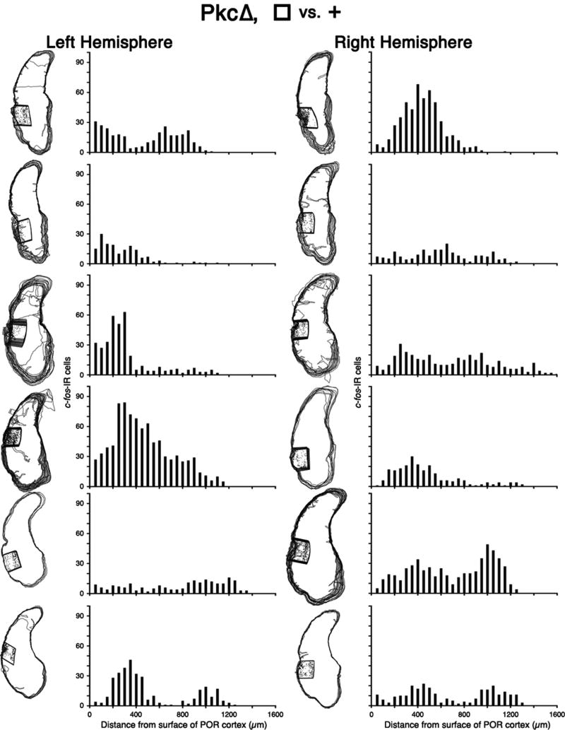
Synaptic plasticity and neural network theories hypothesize that the essential information for advanced cognitive tasks is encoded in specific circuits and neurons within distributed neocortical networks. However, these circuits are incompletely characterized, and we do not know if a specific discrimination is encoded in characteristic circuits among multiple animals. Here, we determined the spatial distribution of active neurons for a circuit that encodes some of the essential information for a cognitive task. We genetically activated protein kinase C pathways in several hundred spatially-grouped glutamatergic and GABAergic neurons in rat postrhinal cortex, a multimodal associative area that is part of a distributed circuit that encodes visual object discriminations. We previously established that this intervention enhances accuracy for specific discriminations. Moreover, the genetically-modified, local circuit in POR cortex encodes some of the essential information, and this local circuit is preferentially activated during performance, as shown by activity-dependent gene imaging. Here, we mapped the positions of the active neurons, which revealed that two image sets are encoded in characteristic and different circuits. While characteristic circuits are known to process sensory information, in sensory areas, this is the first demonstration that characteristic circuits encode specific discriminations, in a multimodal associative area. Further, the circuits encoding the two image sets are intermingled, and likely overlapping, enabling efficient encoding. Consistent with reconsolidation theories, intermingled and overlapping encoding could facilitate formation of associations between related discriminations, including visually similar discriminations or discriminations learned at the same time or place.
Keywords: Activity-dependent gene imaging; Characteristic circuits; Neocortical circuits; Overlapping encoding; Visual discrimination learning.
Copyright © 2017 Elsevier B.V. All rights reserved.
Conflict of interest statement
AIG has equity in Alkermes Inc.
Figures


Similar articles
-
An identified ensemble within a neocortical circuit encodes essential information for genetically-enhanced visual shape learning.Hippocampus. 2019 Aug;29(8):710-725. doi: 10.1002/hipo.23068. Epub 2019 Feb 7. Hippocampus. 2019. PMID: 30734387
-
Identified circuit in rat postrhinal cortex encodes essential information for performing specific visual shape discriminations.Proc Natl Acad Sci U S A. 2010 Aug 10;107(32):14478-83. doi: 10.1073/pnas.0912950107. Epub 2010 Jul 26. Proc Natl Acad Sci U S A. 2010. PMID: 20660720 Free PMC article.
-
CaMKII, MAPK, and CREB are coactivated in identified neurons in a neocortical circuit required for performing visual shape discriminations.Hippocampus. 2012 Dec;22(12):2276-89. doi: 10.1002/hipo.22045. Epub 2012 Jun 27. Hippocampus. 2012. PMID: 22736516
-
From sensation to cognition.Brain. 1998 Jun;121 ( Pt 6):1013-52. doi: 10.1093/brain/121.6.1013. Brain. 1998. PMID: 9648540 Review.
-
From brain synapses to systems for learning and memory: Object recognition, spatial navigation, timed conditioning, and movement control.Brain Res. 2015 Sep 24;1621:270-93. doi: 10.1016/j.brainres.2014.11.018. Epub 2014 Nov 20. Brain Res. 2015. PMID: 25446436 Review.
Cited by
-
Trans-synaptic Neural Circuit-Tracing with Neurotropic Viruses.Neurosci Bull. 2019 Oct;35(5):909-920. doi: 10.1007/s12264-019-00374-9. Epub 2019 Apr 19. Neurosci Bull. 2019. PMID: 31004271 Free PMC article. Review.
References
-
- Squire LR, Kandel ER. Memory. 2. Roberts & Company Publishers; Greenwood Village, CO, US: 2008.
-
- Dudai Y. The Neurobiology of Memory. Oxford Univ. Press, Oxford; England: 1989.
-
- Rumelhart DE, McClelland JL, Group PR. Parallel Distributed Processing. MIT Press; Cambridge MA: 1986.
-
- Dudai Y. The neurobiology of consolidations or how stable is the engram? Annu. Rev. Psychol. 2004;55:51–86. - PubMed
-
- Mumby DG, Pinel JP. Rhinal cortex lesions and object recognition in rats. Behav. Neurosci. 1994;108:11–18. - PubMed
Publication types
MeSH terms
Grants and funding
LinkOut - more resources
Full Text Sources
Other Literature Sources
Medical
